Atlas of Ankle and Foot CT Anatomy
This webpage presents the anatomical structures found on ankle CT.
Click on a link to get
Coronal view - Axial view - Sagittal view
-
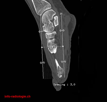
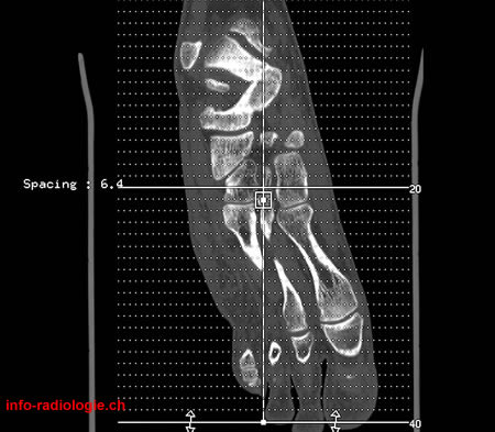
Scout view (coronal reconstructions).
-
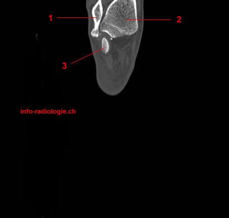
CT-scan of the ankle (coronal reconstruction). Image 2. 1, Fibula. 2, Tibia. 3, Talus.
-
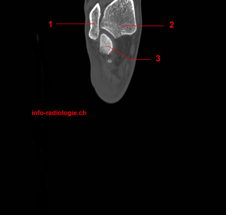
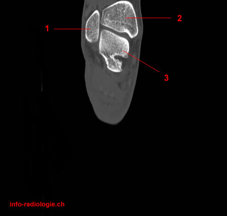
CT-scan of the ankle (coronal reconstruction). Image 3. 1, Fibula. 2, Tibia. 3, Talus.
-
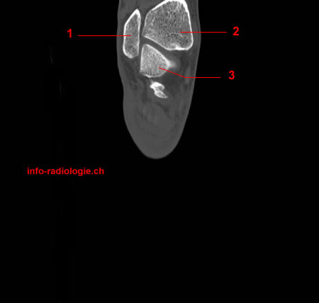
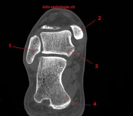
CT-scan of the ankle (coronal reconstruction). Image 4. 1, Fibula. 2, Tibia. 3, Talus.
-
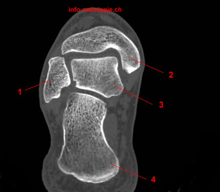
CT-scan of the ankle (coronal reconstruction). Image 5. 1, Fibula. 2, Tibia. 3, Talus.
-
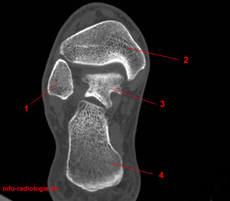
CT-scan of the ankle (coronal reconstruction). Image 6. 1, Fibula. 2, Tibia. 3, Talus. 4, Navicular.
-
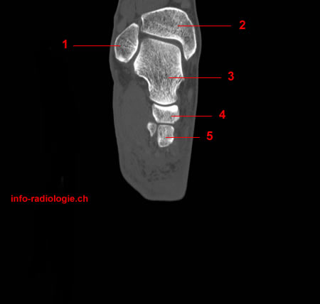
CT-scan of the ankle (coronal reconstruction). Image 7. 1, Fibula. 2, Tibia. 3, Talus. 4, Navicular. 5, Intermediate cuneiform.
-
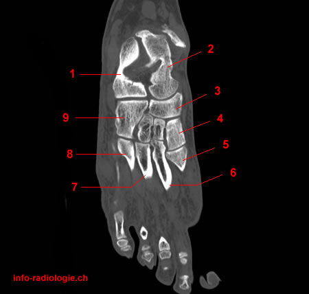
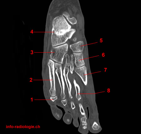
CT-scan of the ankle (coronal reconstruction). Image 8. 1, Fibula. 2, Tibia. 3, Talus. 4, Navicular. 5, Intermediate cuneiform. 6, Medial cuneiform. 7, Base of 2nd metatarsal. 8, Lateral cuneiform.
-
CT-scan of the ankle (coronal reconstruction). Image 9. 1, Fibula. 2, Tibia. 3, Talus. 4, Navicular. 5, Intermediate cuneiform. 6, Medial cuneiform. 7, Base of 2nd metatarsal. 8, Lateral cuneiform.
-
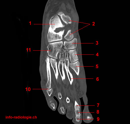
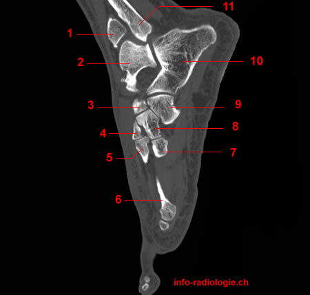
CT-scan of the ankle (coronal reconstruction). Image 10. 1, Fibula. 2, Tibia. 3, Talus. 4, Navicular. 5, Medial cuneiform. 6, Base of 1st metatarsal. 7, Base of 2nd metatarsal. 8, Base of 3rd metatarsal. 9, Lateral cuneiform. 10, Cuboid. 11, Calcaneus.
-
CT-scan of the ankle (coronal reconstruction). Image 11. 1, Fibula. 2, Tibia. 3, Talus. 4, Navicular. 5, Medial cuneiform. 6, Base of 1st metatarsal. 7, Base of 2nd metatarsal. 8, Base of 3rd metatarsal. 9, Lateral cuneiform. 10, Cuboid. 11, Calcaneus.
-
CT-scan of the ankle (coronal reconstruction). Image 12. 1, Calcaneus. 2, Talus. 3, Navicular. 4, Medial cuneiform. 5, Base of 1st metatarsal. 6, 2nd metatarsal. 7, 3rd metatarsal. 8, Base of 4th metatarsal. 9, Cuboid.
-
CT-scan of the ankle (coronal reconstruction). Image 13. 1, Calcaneus. 2, Talus. 3, Navicular. 4, Medial cuneiform. 5, Base of 1st metatarsal. 6, 2nd metatarsal. 7, 2nd proximal phalanx. 8, 2nd middle phalanx. 9, 2nd distal phalanx. 10, 5th metatarsophalangeal joint. 11, Cuboid.
-
CT-scan of the ankle (coronal reconstruction). Image 14. 1, Base of 5th proximal phalanx. 2, 5th metatarsal. 3, Cuboid. 4, Calcaneus. 5, Navicular. 6, Medial cuneiform. 7, Base of 1st metatarsal. 8, 2nd metatarsal.
-
CT-scan of the ankle (coronal reconstruction). Image 15. 1, 5th metatarsal. 2, Cuboid. 3, Calcaneus. 4, Navicular. 5, Medial cuneiform. 6, 1st metatarsal. 7, 2nd metatarsophalangeal joint. 8, Distal phalanx. 9, base of 3rd proximal phalanx. 10, base of 4th proximal phalanx.
-
CT-scan of the ankle (coronal reconstruction). Image 16. 1, Base of 5th metatarsal. 2, Cuboid. 3, Calcaneus. 4, Navicular. 5, Medial cuneiform. 6, 1st metatarsal. 7, 1st proximal phalanx. 8, 1st distal phalanx. 9, base of 2nd proximal phalanx. 10, 4th metatarsal.
-
CT-scan of the ankle (coronal reconstruction). Image 17. 1, Base of 5th metatarsal. 2, Calcaneus. 3, Medial cuneiform. 4, 1st metatarsal. 5, 1st metatarsophalangeal joint. 6, 1st distal phalanx.
-
CT-scan of the ankle (coronal reconstruction). Image 18. 1, Base of 5th metatarsal. 2, Cuboid. 3, Calcaneus. 4, Medial cuneiform. 5, 1st metatarsal. 6, 1st proximal phalanx. 7, 1st distal phalanx.
-
CT-scan of the ankle (coronal reconstruction). Image 19. 1, Calcaneus. 2, 1st metatarsophalangeal joint.
-
CT-scan of the ankle (coronal reconstruction). Image 20. 1, Calcaneus. 2, 1st metatarsal. 3, 1st proximal phalanx.
-
Scout view (Axial reconstruction)
-
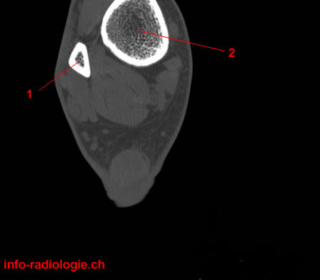
CT-scan of the ankle (axial reconstruction). Image 1. 1, Fibula. 2, Tibia.
-
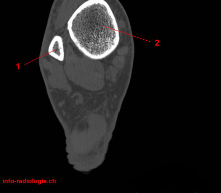
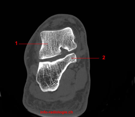
CT-scan of the ankle (axial reconstruction). Image 2. 1, Fibula. 2, Tibia.
-
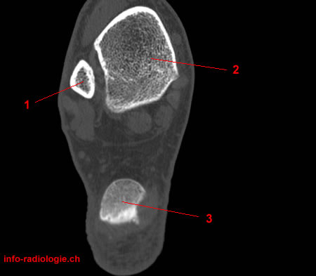
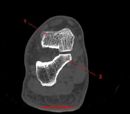
CT-scan of the ankle (axial reconstruction). Image 3. 1, Fibula. 2, Tibia. 3, Calcaneus.
-
CT-scan of the ankle (axial reconstruction). Image 4. 1, Fibula. 2, Tibia. 3, Calcaneus.
-
CT-scan of the ankle (axial reconstruction). Image 5. 1, Fibula. 2, Tibia. 3, Talus. 4, Calcaneus.
-
CT-scan of the ankle (axial reconstruction). Image 6. 1, Fibula. 2, Tibia. 3, Talus. 4, Calcaneus.
-
CT-scan of the ankle (axial reconstruction). Image 7. 1, Fibula. 2, Tibia. 3, Talus. 4, Calcaneus.
-
CT-scan of the ankle (axial reconstruction). Image 8. 1, Fibula. 2, Tibia (medial malleolus). 3, Talus. 4, Calcaneus.
-
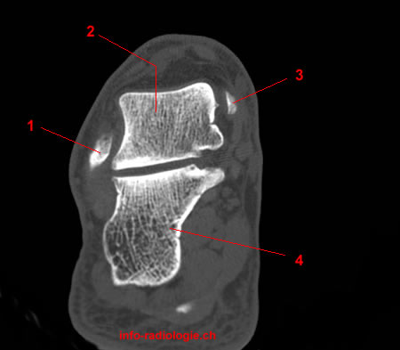
CT-scan of the ankle (axial reconstruction). Image 9. 1, Fibula. 2, Talus. 3, Tibia (medial malleolus). 4, Calcaneus.
-
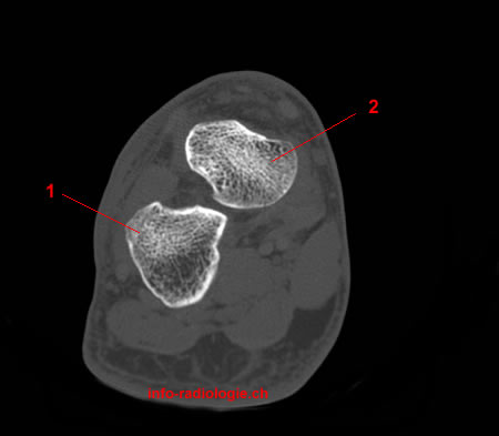
CT-scan of the ankle (axial reconstruction). Image 10. 1, Talus. 2, Calcaneus.
-
CT-scan of the ankle (axial reconstruction). Image 11. 1, Talus. 2, Calcaneus.
-
CT-scan of the ankle (axial reconstruction). Image 12. 1, Talus. 2, Calcaneus.
-
CT-scan of the ankle (axial reconstruction). Image 13. 1, Calcaneus. 2, Talus.
-
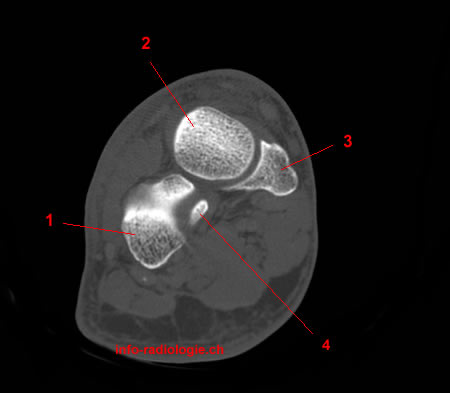
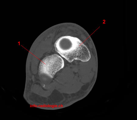
CT-scan of the ankle (axial reconstruction). Image 14. 1, Calcaneus. 2, Talus. 3, Navicular. 4, Cuboid.
-
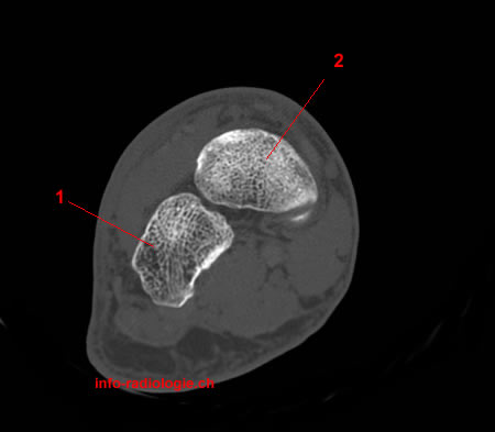
CT-scan of the ankle (axial reconstruction). Image 15. 1, Cuboid 2, Navicular.
-
CT-scan of the ankle (axial reconstruction). Image 16. 1, Cuboid 2, Navicular.
-
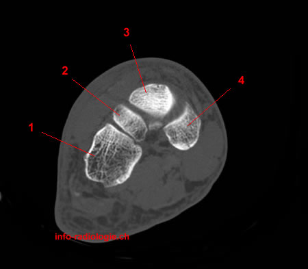
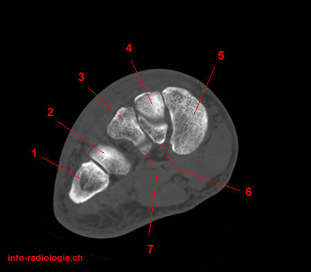
CT-scan of the ankle (axial reconstruction). Image 17. 1, Cuboid. 2, Lateral cuneiform. 3, Navicular. 4, Medial cuneiform.
-
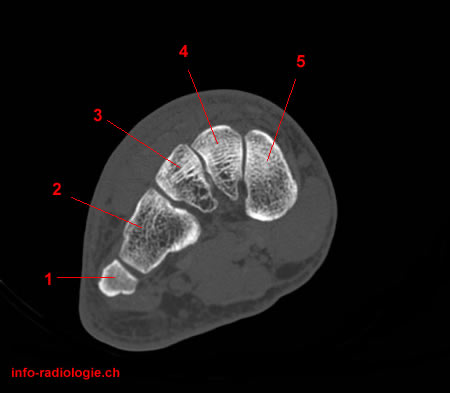
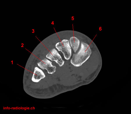
CT-scan of the ankle (axial reconstruction). Image 18. 1, 5th metatarsal. 2, Cuboid. 3, Lateral cuneiform. 4, Intermediate cuneiform. 5, Medial cuneiform.
-
CT-scan of the ankle (axial reconstruction). Image 19. 1, 5th metatarsal. 2, Cuboid. 3, Lateral cuneiform. 4, Intermediate cuneiform. 5, Medial cuneiform.
-
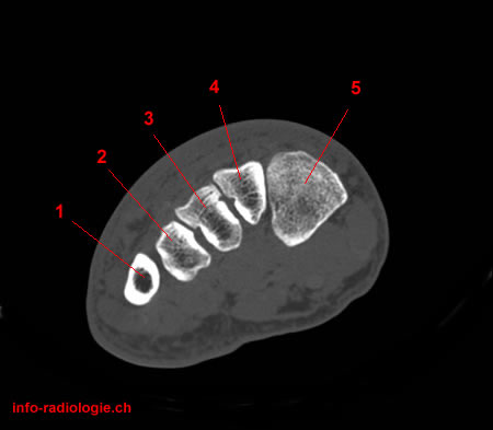
CT-scan of the ankle (axial reconstruction). Image 20. 1, 5th metatarsal. 2, 4th metatarsal. 3, Lateral cuneiform. 4, Intermediate cuneiform. 5, Medial cuneiform. 6, 2nd metatarsal. 7, 3rd metatarsal.
-
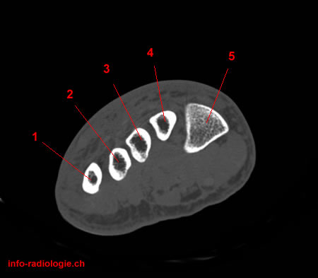
CT-scan of the ankle (axial reconstruction). Image 21. 1, 5th metatarsal. 2, 4th metatarsal. 3, 3rd metatarsal. 4, 2nd metatarsal. 5, Medial cuneiform. 6, 1st metatarsal.
-
CT-scan of the ankle (axial reconstruction). Image 22. 1, 5th metatarsal. 2, 4th metatarsal. 3, 3rd metatarsal. 4, 2nd metatarsal. 5, 1st metatarsal.
-
CT-scan of the ankle (axial reconstruction). Image 23. 1, 5th metatarsal. 2, 4th metatarsal. 3, 3rd metatarsal. 4, 2nd metatarsal. 5, 1st metatarsal.
-
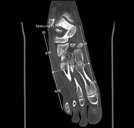
Scout view (sagittal reconstruction)
-
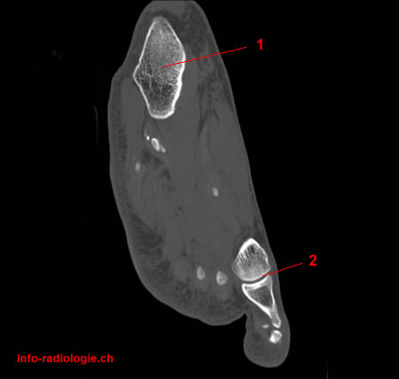
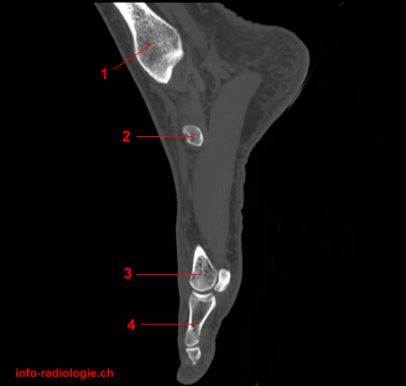
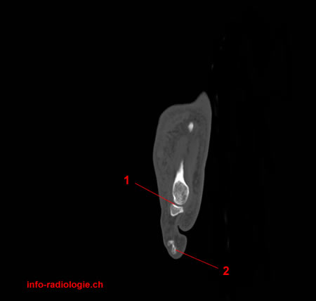
CT-scan of the ankle (sagittal reconstruction). Image 2. 1, Tibia (medial malleolus). 2, 1st Metatarsophalangeal joint.
-
CT-scan of the ankle (sagittal reconstruction). Image 3. 1, Tibia (medial malleolus). 2, Navicular. 3, 1st metatarsal. 4, Proximal phalanx.
-
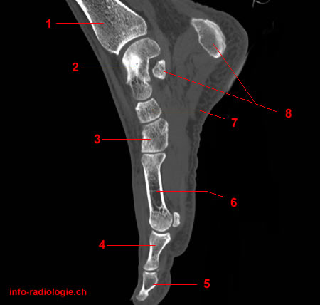
CT-scan of the ankle (sagittal reconstruction). Image 4. 1, Tibia. 2, Navicular. 3, 1st metatarsal. 4, Distal phalanx. 5, Proximal phalanx. 6, Medial cuneiform. 7, Calcaneus. 8, Talus.
-
CT-scan of the ankle (sagittal reconstruction). Image 5. 1, Tibia. 2, Talus. 3, Medial cuneiform. 4, Proximal phalanx. 5, Distal phalanx. 6, 1st metatarsal. 7, Navicular. 8, Calcaneus.
-
CT-scan of the ankle (sagittal reconstruction). Image 6. 1, Tibia. 2, Talus. 3, Medial cuneiform. 4, Proximal phalanx. 5, Distal phalanx. 6, 1st metatarsal. 7, Navicular. 8, Calcaneus.
-
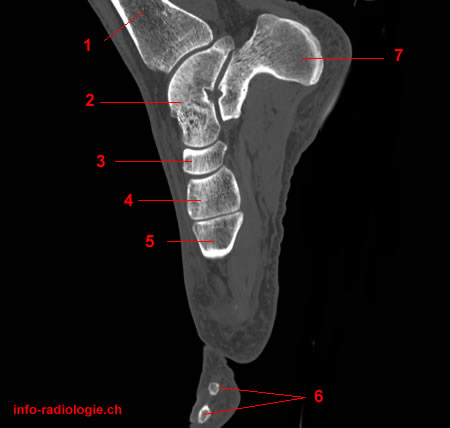
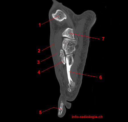
CT-scan of the ankle (sagittal reconstruction). Image 7. 1, Tibia. 2, Talus. 3, Navicular. 4, Medial cuneiform. 5, Base of 1st metatarsal. 6, 1st distal phalanx. 7, Calcaneus.
-
CT-scan of the ankle (sagittal reconstruction). Image 8. 1, Tibia. 2, Talus. 3, Navicular. 4, Medial cuneiform. 5, Base of 1st metatarsal. 6, Calcaneus.
-
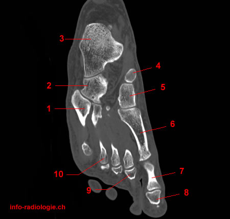
CT-scan of the ankle (sagittal reconstruction). Image 9. 1, Tibia. 2, Talus. 3, Navicular. 4, Medial cuneiform. 5, Base of 1st metatarsal. 6, 2nd metatarsal. 7, 2nd proximal phalanx. 8, Intermediate cuneiform. 9, Calcaneus.
-
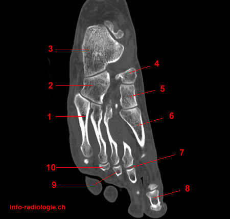
CT-scan of the ankle (sagittal reconstruction). Image 10. 1, Tibia. 2, Talus. 3, Navicular. 4, Intermediate cuneiform. 5, 2nd metatarsal. 6, Metatarsophalangeal joint. 7, Distal interphalangeal joint. 8, Proximal interphalangeal joint. 9, Cuboid. 10, Calcaneus.
-
CT-scan of the ankle (sagittal reconstruction). Image 11. 1, Tibia. 2, Talus. 3, Navicular. 4, Intermediate cuneiform. 5, 2nd metatarsal. 6, 2nd Middle phalanx. 7, Lateral cuneiform. 8, Cuboid. 9, Calcaneus.
-
CT-scan of the ankle (sagittal reconstruction). Image 12. 1, Tibia. 2, Talus. 3, Navicular. 4, Intermediate cuneiform. 5, base of 2nd metatarsal. 6, 3rd metatarsal. 7, base of 3rd metatarsal. 8, Lateral cuneiform. 9, Cuboid. 10, Calcaneus. 11, Fibula.
-
CT-scan of the ankle (sagittal reconstruction). Image 13. 1, Tibia. 2, Talus. 3, Lateral cuneiform. 4, 3rd metatarsal. 5, 3rd Metatarsophalangeal joint. 6, Cuboid. 7, Calcaneus. 8, Fibula.
-
CT-scan of the ankle (sagittal reconstruction). Image 14. 1, Fibula. 2, Cuboid. 3, Lateral cuneiform. 4, Base of 3rd metatarsal. 5, 3rd proximal interphalangeal joint. 6, base of 4th metatarsal. 7, Calcaneus.
-
CT-scan of the ankle (sagittal reconstruction). Image 15. 1, Fibula. 2, Cuboid. 3, Lateral cuneiform. 4, Base du 3rd metatarsal. 5, 3rd distal interphalangeal joint. 6, 4th metatarsal. 7, Calcaneus.
-
CT-scan of the ankle (sagittal reconstruction). Image 16. 1, Fibula. 2, Cuboid. 3, 4th metatarsal. 4, 4th Metatarsophalangeal joint. 5, base of 5th metatarsal.
-
CT-scan of the ankle (sagittal reconstruction). Image 17. 1, Cuboid. 2, Base of 4th metatarsal. 3, 4th Proximal interphalangeal joint. 4, 4th Distal phalanx. 5, base of 5th metatarsal.
-
CT-scan of the ankle (sagittal reconstruction). Image 18. 1, 5th metatarsal. 2, 4th Proximal interphalangeal joint. 3, 4th Distal interphalangeal joint.
-
CT-scan of the ankle (sagittal reconstruction). Image 19. 1, 5th metatarsal.
-
CT-scan of the ankle (sagittal reconstruction). Image 20. 1, 5th Metatarsophalangeal joint. 2, Distal phalanx.