This webpage presents the anatomical structures found on elbow radiographs.
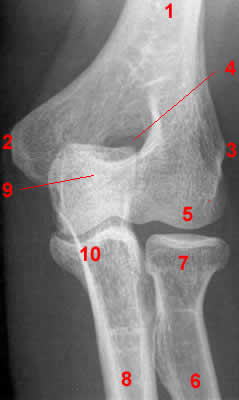 1, Humerus. 2, Medial epicondyle. 3, Lateral epicondyle. 4, Olecranon fossa. 5, Capitulum. 6, Radius. 7, Radial head. 8, Ulna. 9, Olecranon. 10, Coronoid process.
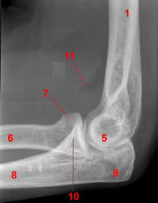 1, Humerus. 5, capitulum. 6, Radius. 7, Radial head. 8, Ulna. 9, Olecranon. 10, Coronoid process of ulna. 11, Lateral radiograph shows a positive fat pad sign in a patient with a nondisplaced fracture of the radial head. The anterior lucency represents the elevated anterior fat pad sign.