-
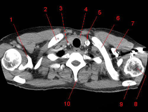
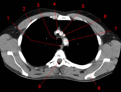
Image 1. CT Anatomy of the chest, axial reconstruction. 1, Coracoid Process (scapula). 2, Clavicle (right side). 3, Right common carotid artery. 4, Thyroid. 5, Internal jugular vein (left side). 6, Clavicle (left side). 7, Subclavian vein (left side). 8, Humeral head (left side). 9, Spine of scapula. 10, Spinous process.
-
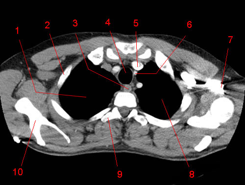
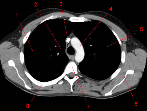
Image 2. CT Anatomy of the chest, axial reconstruction. 1, Humeral head (right side). 2, Oesophagus. 3, Trachea. 4, Left subclavian vein. 5, Spine of scapula. 6, Neck of the scapula.
-
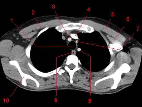
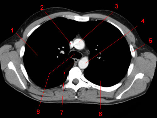
Image 3. CT Anatomy of the chest, axial reconstruction. 1, Lung (right side) 2, Rib. 3, Oesophagus. 4, Trachea. 5, Brachiocephalic vein (left side). 6, Common carotid artery (left side). 7, Axillary vein (left side). 8, Lung (left side). 9, Transverse process. 10, Scapula.
-
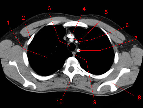
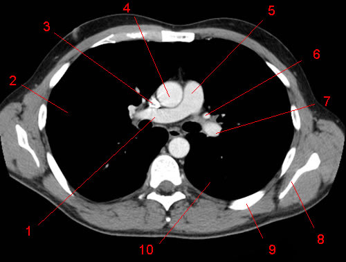
Image 4. CT Anatomy of the chest, axial reconstruction. 1, Trachea. 2, Rib. 3, Brachiocephalic artery. 4, Brachiocephalic vein (left side). 5, Pectoralis major muscle. 6, Pectoralis minor muscle. 7, Common carotid artery (left side). 8, Subclavian artery (left side). 9, Oesophagus. 10, Scapula.
-
Image 5. CT Anatomy of the chest, axial reconstruction. 1, Lung (right side) 2, Rib. 3, Trachea. 4, Brachiocephalic vein (left side). 5, Brachiocephalic artery. 6, Common carotid artery (left side). 7, Subclavian artery (left side). 8, Scapula. 9, Oesophagus. 10, Spinous process.
-
Image 6. CT Anatomy of the chest, axial reconstruction. 1, Oesophagus. 2, Rib. 3, Trachea. 4, Superior vena cava. 5, Brachiocephalic artery. 6, Common carotid artery (left side). 7, Aorta. 8, Scapula. 9, Vertebral foramen.
-
Image 7. CT Anatomy of the chest, axial reconstruction. 1, Lung (right side) 2, Trachea. 3, Superior vena cava. 4, Aortic arch. 5, Lung (left side). 6, Scapula. 7, Vertebral body. 8, Rib.
-
Image 8. CT Anatomy of the chest, axial reconstruction. 1, Lung (right side) 2, Superior vena cava. 3, Ascending thoracic aorta. 4, Descending thoracic aorta. 5, Rib. 6, Lung (left side). 7, Oesophagus. 8, Trachea.
-
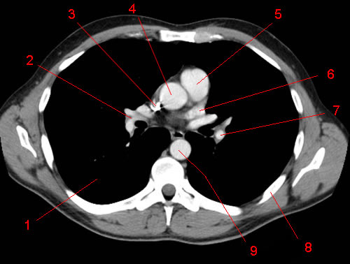
Image 9. CT Anatomy of the chest, axial reconstruction. 1, Lung (right side) 2, Pulmonary vein (right side). 3, Superior vena cava. 4, Ascending thoracic aorta. 5, Pulmonary artery (left side). 6, Pulmonary vein (left side). 7, Lung (left side). 8, Scapula. 9, Vertebral foramen. 10, Rib.
-
Image 10. CT Anatomy of the chest, axial reconstruction. 1, Pulmonary artery (right side). 2, Lung (right side) 3, Superior vena cava. 4, Ascending thoracic aorta. 5, Pulmonary trunk. 6, Pulmonary vein (left side). 7, Pulmonary artery (left side). 8, Scapula. 9, Rib 10, Lung (left side).
-
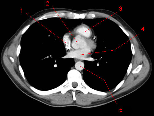
Image 11. CT Anatomy of the chest, axial reconstruction. 1, Lung (right side) 2, Pulmonary artery (right side). 3, Superior vena cava. 4, Ascending thoracic aorta. 5, Pulmonary artery root. 6, Pulmonary vein (left side). 7, Pulmonary artery (left side). 8, Rib. 9, Descending thoracic aorta
-
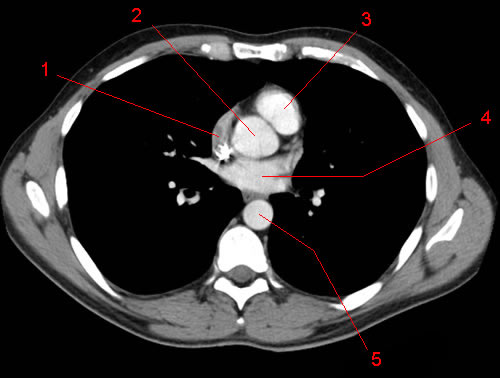
Image 12. CT Anatomy of the chest, axial reconstruction. 1, Right atrium. 2, Aortic root. 3, Pulmonary artery root. 4, Left atrium. 5, Descending thoracic aorta.
-
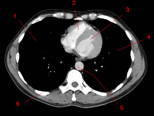
Image 13. CT Anatomy of the chest, axial reconstruction. 1, Right atrium. 2, Aortic root. 3, Ventricle (right side) 4, Left atrium. 5, Descending thoracic aorta
-
Image 14. CT Anatomy of the chest, axial reconstruction. 1, Lung (right side) 2, Right atrium. 3, Ventricle (right side) 4, Ventricle (left side). 5, Lung (left side). 6, Descending thoracic aorta.
-
Image 15. CT Anatomy of the chest, axial reconstruction. 1, Lung (right side) 2, Ventricle (right side) 3, Ventricle (left side). 4, Lung (left side). 5, Descending thoracic aorta. 6, Rib.
-
Image 16. CT Anatomy of the chest, axial reconstruction. 1, Oesophagus. 2, Lung (right side) 3, Ventricle (right side) 4, Ventricle (left side). 5, Lung (left side). 6, Descending thoracic aorta. 7, Spinous process.
-
Image 17. CT Anatomy of the chest, axial reconstruction. 1, Lung (right side) 2, Inferior vena cava. 3, Ventricle (right side) 4, Ventricle (left side). 5, Oesophagus. 6, Lung (left side). 7, Descending thoracic aorta. 8, Vertebral foramen.
-
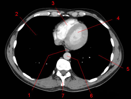
Image 18. CT Anatomy of the chest, axial reconstruction. 1, Lung (right side) 2, Liver. 3, Inferior vena cava. 4, Ventricle (right side) 5, Ventricle (left side). 6, Oesophagus. 7, Lung (left side). 8, Descending thoracic aorta. 9, Vertebral body.
-
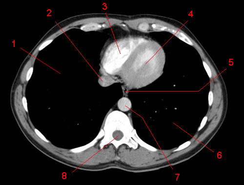
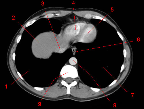
Image 19. CT Anatomy of the chest, axial reconstruction. 1, Inferior lobe of right lung. 2, Liver. 3, Inferior vena cava. 4, Oesophagus. 5, Lung (left side) 6, Aorta.
-
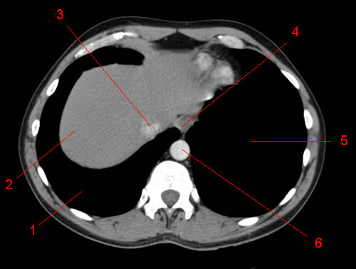
Image 20 of 20. CT Anatomy of the chest, axial reconstruction. 1, Inferior lobe of right lung. 2, Liver. 3, Inferior vena cava. 4, Inferior lobe of left lung. 5, Aorta.