-
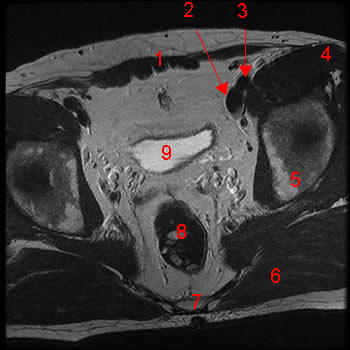
MRI of the male pelvis: T1-weighted, axial view. Image 1. 1, rectus abdominis m. 2, external iliac v. 3, external iliac a. 4, sartorius m. 5, ilium 6, gluteus maximus m. 7, sacrum 8, rectum 9, bladder
-
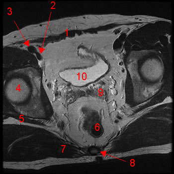
MRI of the male pelvis: T1-weighted, axial view. Image 2. 1, rectus abdominis m. 2, external iliac v. 3, external iliac a. 4, femoral head 5, piriformis m. 6, rectum 7, gluteus maximus m. 8, sacrum 9, seminal vesicle 10, bladder
-
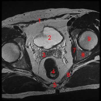
MRI of the male pelvis: T1-weighted, axial view. Image 3. 1, rectus abdominis m. 2, bladder 3, seminal vesicle 4, rectum 5, coccyx 6, coccygeus m. 7, obturator internus m. 8, ischium 9, femoral head
-
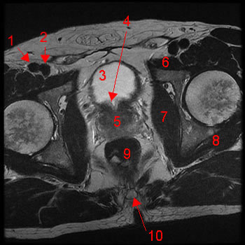
MRI of the male pelvis: T1-weighted, axial view. Image 4. 1, femoral a. 2, femoral v. 3, bladder 4, pectineus m. 5, iliopsoas m. 6, sartorius m. 7, femoral head 8, obturator internus m. 9, gemellus (superior, inferior) m. 10, coccygeus m. 11, rectum
-
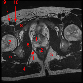
MRI of the male pelvis: T1-weighted, axial view. Image 5. 1, femoral a. 2, femoral v. 3, bladder 4, bladder neck 5, prostate 6, pectineus m. 7, obturator internus m. 8, gemellus (superior, inferior) m. 9, rectum 10,coccyx
-
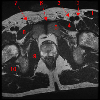
MRI of the male pelvis: T1-weighted, axial view. Image 6. 1, levator ani m. 2, rectum 3, ischiorectal fossa 4, gluteus maximus m. 5, gemellus (superior, inferior) m. 6, obturator internus m. 7, femoral head 8, pectineus m. 9, femoral a. 10,femoral v. 11, prostate
-
MRI of the male pelvis: T1-weighted, axial view. Image 7. 1, sartorius m. 2, femoral a. 3,femoral v. 4, left pectineus m. 5, pubis 6, symphysis pubis 7, spermatic cord 8, right pectineus m. 9, obturator internus m. 10, gemellus inferior m.
-
MRI of the male pelvis: T1-weighted, axial view. Image 8. 1, corpus cavernosum 2, corpus spongiosum (bulb of the penis) 3, ramus ischium 4, ischiocavernosus m. 5, anal canal 6, sphincter ani externus m. 7, gluteus maximus m.
-
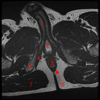
MRI of the male pelvis: T1-weighted, coronal view. Image 9. 1, rectus abdominis m. 2, bladder 3, pubis 4, ischium 5, testis 6, corpus cavernosum
-
MRI of the male pelvis: T1-weighted, coronal view. Image 10. 1, rectus abdominis m. 2,pubis 3, bladder 4, prostate 5, rectum 6, sigmoid colon
-
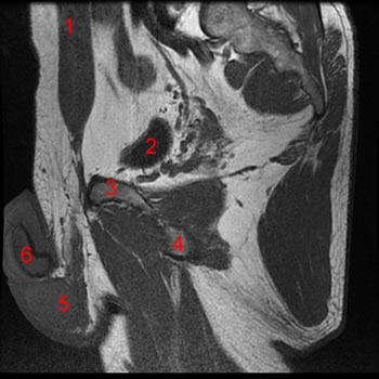
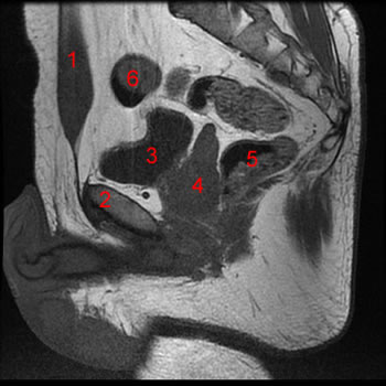
MRI of the male pelvis: T1-weighted, coronal view. Image 11. 1, rectus abdominis m. 2,median umbilical ligament 3, bladder 4, symphysis pubis 5, prostate 6, seminal vesicle 7,rectum 8, corpus cavernosum
-
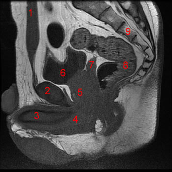
MRI of the male pelvis: T1-weighted, coronal view. Image 12 of 12. 1, rectus abdominis m. 2, symphysis pubis 3, corpus cavernosum 4, corpus spongiosum 5, prostate 6, bladder 7,seminal vesicle 8, rectum 9, sacrum