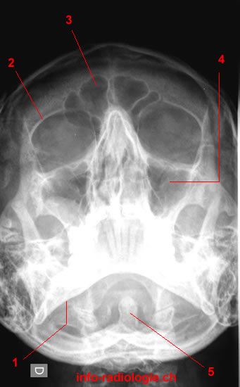
Radiography of the paranasal sinuses. Image 1 1, mandible. 2, Upper edge of the orbit. 3, Frontal sinus. 4, Maxillary sinus. 5, Odontoid process.
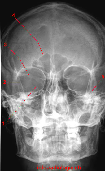
Radiography of the paranasal sinuses. Image 2 1, Superior orbital fissure. 2, Greater wing of sphenoid. 3, Lesser wing of sphenoid. 4, Frontal sinus. 5, Petrous bone.
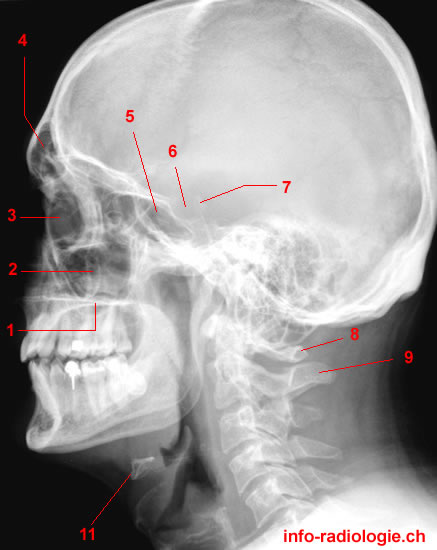
Radiography of the paranasal sinuses. Image 3 of 3. 1, Hard palate. 2, Maxillary sinus. 3, Orbit. 4, Frontal sinus. 5, Sinus sphénoïdal. 6, Pituitary fossa. 7, Posterior clinoid process. 8, Spinous process (Atlas). 9, Spinous process (Axis).