-
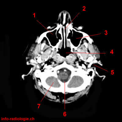
Image 1, cerebral CT, axial reconstruction. 1, Maxillary sinus (Right side). 2, Nasal septum. 3, Maxillary sinus (left side). 4, Nasopharynx. 5, External auditory meatus. 6, Foramen magnum. 7, Cerebellum.
-
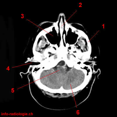
Image 2, cerebral CT, axial reconstruction. 1, Zygomatic arch. 2, Nasal septum. 3, Maxillary sinus (right side). 4, External auditory meatus. 5, Medulla. 6, Cerebellum.
-
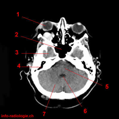
Image 3, cerebral CT, axial reconstruction. 1, Globe. 2, Sphenoidal sinus. 3, Temporal lobe (right side). 4, Mastoid cells. 5, Pons. 6, Fourth ventricle. 7, cerebellar hemisphere.
-
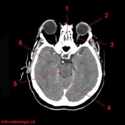
Image 4, cerebral CT, axial reconstruction. 1, Ethmoidal cells. 2, Globe. 3, Optic nerve. 4, Vermis. 5, Midbrain. 6, Temporal lobe.
-
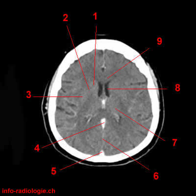
Image 5, cerebral CT, axial reconstruction. 1, Caudate nucleus. 2, Anterior limb, internal capsule. 3, Lenticular nucleus. 4, Inferior sagittal sinus. 5, Superior sagittal sinus. 6, Interhemispheric fissure / Falx cerebri. 7, Thalamus. 8, Lateral ventricle. 9, Corpus callosum.
-
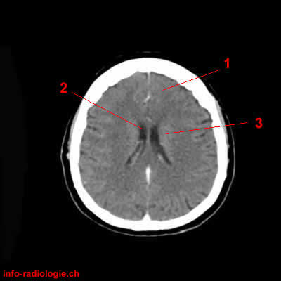
Image 6, cerebral CT, axial reconstruction. 1, Superior frontal gyrus. 2, Lateral ventricle. 3, Caudate nucleus.
-
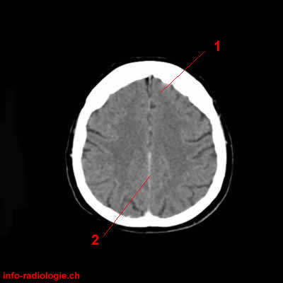
Image 7, cerebral CT, axial reconstruction. 1, Superior frontal gyrus. 2, Interhemispheric fissure / Falx cerebri.
-
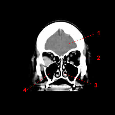
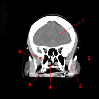
Image 8, Cerebral CT, coronal reconstruction. 1, Frontal lobe. 2, Lateral rectus muscle. 3, Nasal turbinate. 4, Maxillary sinus.
-
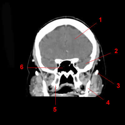
Image 9, Cerebral CT, coronal reconstruction. 1, Frontal lobe. 2, Zygomatic arch. 3, Mandible. 4, Nasal turbinate. 5, Alveolar arch. 6, Maxillary sinus (right side).
-
Image 10, Cerebral CT, coronal reconstruction. 1, Frontal lobe. 2, Temporal lobe. 3, Zygomatic arch. 4, Mandible. 5, Alveolar arch. 6, Sphenoid sinus.
-
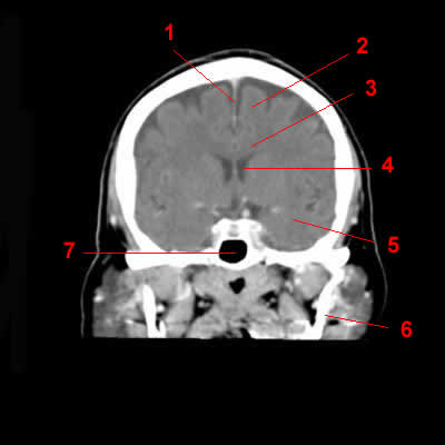
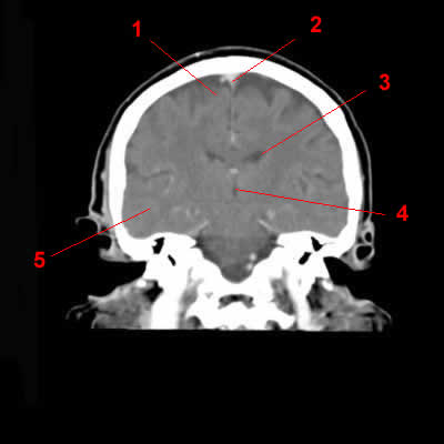
Image 11, Cerebral CT, coronal reconstruction. 1, Interhemispheric fissure / Falx cerebri. 2, Frontal lobe. 3, Corpus callosum. 4, Lateral ventricle. 5, Temporal lobe. 6, Mandible. 7, Sphenoid sinus.
-
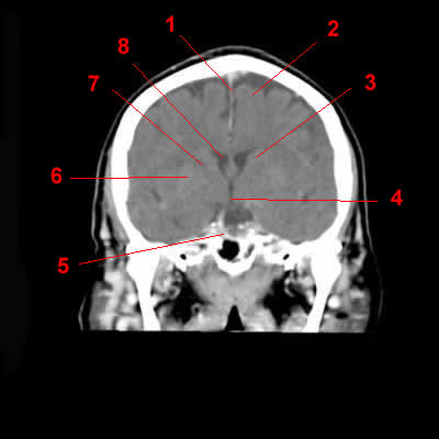
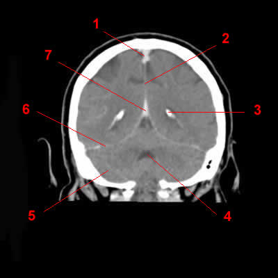
Image 12, Cerebral CT, coronal reconstruction. 1, Interhemispheric fissure / Falx cerebri. 2, Superior frontal gyrus. 3, Caudate nucleus. 4, Third ventricule. 5, Basilar artery. 6, Lenticular nucleus. 7, Internal capsule. 8, Lateral ventricle.
-
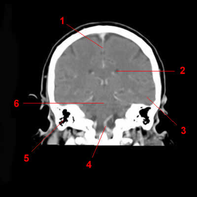
Image 13, Cerebral CT, coronal reconstruction. 1, Superior frontal gyrus. 2, Superior sagittal sinus. 3, Lateral ventricle. 4, Third ventricle. 5, Temporo-occipital Gyri.
-
Image 14, Cerebral CT, coronal reconstruction. 1, Interhemispheric fissure / Falx cerebri. 2, Lateral ventricle. 3, Temporo-occipital Gyri (left side). 4, Vertebral artery. 5, Mastoid sinuses (right side). 6, Cerebral trunk.
-
Image 15, Cerebral CT, coronal reconstruction. 1, Superior sagittal sinus. 2, Interhemispheric fissure / Falx cerebri. 3, Choroid plexus. 4, Fourth ventricle. 5, Cerebellum. 6, Tentorium cerebelli. 7, Straight sinus.
-
Image 16, Cerebral CT, sagittal reconstruction. 1, Globe. 2, Frontal lobe. 3, Sylvian fissure. 4, Occipital lobe. 5, Tentorium cerebelli. 6, Cerebellar hemisphere. 7, Parahippocampus.
-
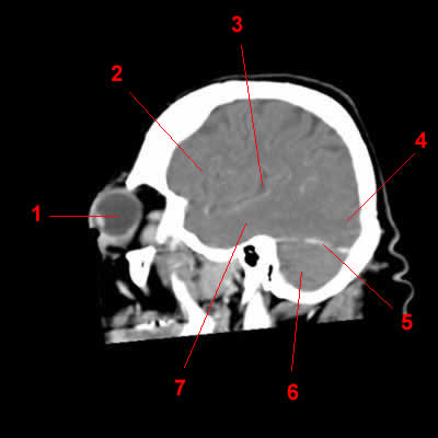
Image 17, Cerebral CT, sagittal reconstruction. 1, Maxillary sinus. 2, Globe. 3, Frontal lobe. 4, Lateral ventricle / Choroid plexus. 5, Temporo-occipital Gyri. 6, Tentorium cerebelli. 7, cerebellar hemisphere.
-
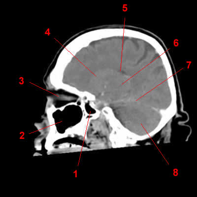
Image 18, Cerebral CT, sagittal reconstruction. 1, Sphenoidal sinus. 2, Maxillary sinus. 3, Optic nerve. 4, Caudate nucleus. 5, Lateral ventricle. 6, Thalamus. 7, Tentorium cerebelli. 8, Cerebellar hemisphere.
-
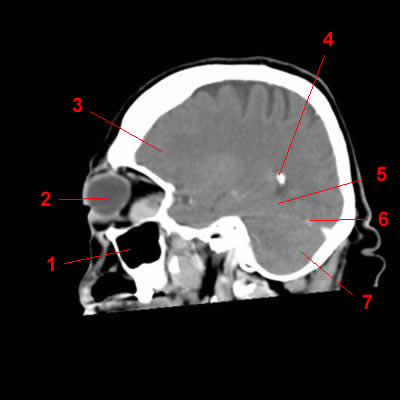
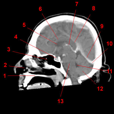
Image 19, Cerebral CT, sagittal reconstruction. 1, Bony palate. 2, Nasal fossa. 3, Sphenoidal sinus. 4, Turcic sellae. 5, Lateral ventricle. 6, Corpus callosum. 7, Internal cerebral vein. 8, Inferior sagittal sinus. 9, Straight sinus. 10, Confluens sinuum. 11, Fourth ventricle. 12, cerebellar hemisphere. 13, Pons
-
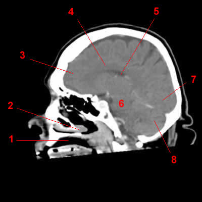
Image 20 of 20, Cerebral CT, sagittal reconstruction. 1, Oropharynx. 2, Nasal turbinate. 3, Frontal lobe. 4, Corpus callosum. 5, Lateral ventricle. 6, Cerebral trunk. 7, Occipital lobe. 8, Cerebellar hemisphere.