Cervical Spine Radiographs

This photo gallery presents the anatomical structures found on cervical spine radiographs.

Image 1. Cervical Spine X-ray: AP view.
1, Clavicle. 2, 1st rib. 3, Trachea. 4, Spinous process of C7. 5, Vertebral body of C5. 6, Uncinate process.

Radiographie du rachis cervical: Image 1
  • Image 1. Cervical Spine X-ray: AP view.
    1, Clavicle. 2, 1st rib. 3, Trachea. 4, Spinous process of C7. 5, Vertebral body of C5. 6, Uncinate process.

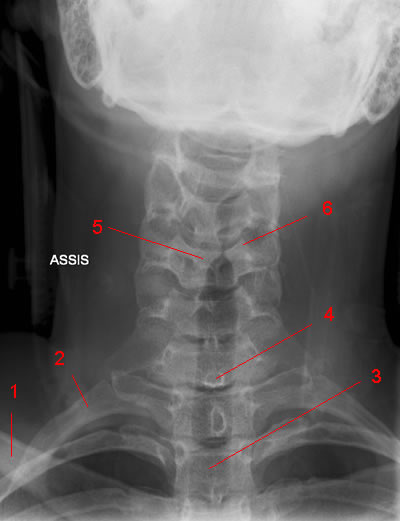
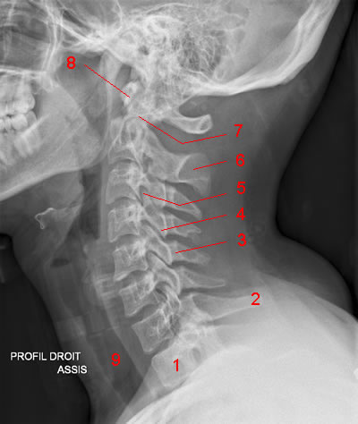
  • Image 2. Cervical Spine X-ray: Lateral view.
    1, Vertebral body (TH1). 2, Spinous process of C7. 3, Lamina. 4, Inferior articular process. 5, Superior articular process. 6, Spinous process of C2. 7, Odontoid process. 8, Anterior arch of C1 (Atlas). 9, Trachea.

  • Image 3. C1 and odontoid process: AP view (Mouth wide open). 1, Transverse process of C1. 2, Lateral mass of C1. 3, Odontoid process. 4, Inferior articular process of C1. 5, Superior articular process of C2.

  • Image 4. Cervical Spine X-ray, 3/4 (Left Neural Foramina).
    1, Rib. 2, Clavicle. 3, Intervertebral foramen. 4, Pedicle. 5, Trachea.

  • Image 5 of 5. Cervical Spine X-ray, 3/4 (Right Neural Foramina). 1, Clavicle. 2, Rib. 3, Lamina. 4, Intervertebral foramen. 5, Uncinate process. 6, Pedicle.