-
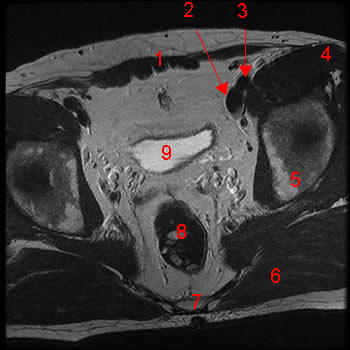
MRI of the female pelvis: T1-weighted, axial view. Image 1. 1, Rectus abdominis m. 2, external iliac vein 3, external iliac artery 4, right ovary 5, uterus 6, left ovary 7, ilium 8, rectum 9, sacrum
-
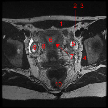
MRI of the female pelvis: T1-weighted, axial view. Image 2. 1, Rectus abdominis m. 2, external iliac vein 3, external iliac artery 4, obturator internus m. 5, right ovary 6, endometrium 7, junctional zone 8, myometrium 9, left ovary 10, rectum
-
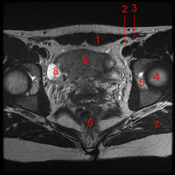
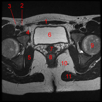
MRI of the female pelvis: T1-weighted, axial view. Image 3. 1, Rectus abdominis m. 2, external iliac vein 3, external iliac artery 4, femoral head 5, acetabulum 6, rectum 7, gluteus maximus 8, right ovary
-
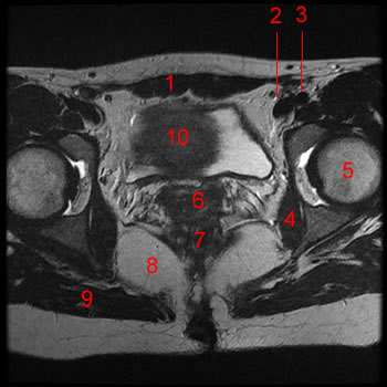
MRI of the female pelvis: T1-weighted, axial view. Image 4. 1, Rectus abdominis m. 2, external iliac vein 3, external iliac artery 4, obturator internus m. 5, head of the femur 6, endocervical canal 7, rectum 8, ischiorectal fossa 9, gluteus maximus 10, uterus
-
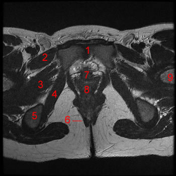
MRI of the female pelvis: T1-weighted, axial view. Image 5. 1, Rectus abdominis m. 2, bladder 3, sartorius m. 4, iliopsoas m. 5, pectineus m 6, pubis 7, head of the femur 8, ilium 9, obturator internus m. 10, gemellus sup. m. 11, gluteus maximus m. 12, ischiorectal fossa 13, vagina 14, rectum
-
MRI of the female pelvis: T1-weighted, axial view. Image 6. 1, Rectus abdominis m. 2, femoral vein 3, femoral artery 4, pectineus muscle 5, obturator internus m.6, bladder 7, vagina 8, anal canal 9, head of the femur 10, ischiorectal fossa 11, gluteus maximus m.
-
MRI of the female pelvis: T1-weighted, axial view. Image 7. 1, Rectus abdominis m.
2, bladder
3, obturator internus m.
4, acetabular fossa
5, rectum
6, vagina
-
MRI of the female pelvis: T1-weighted, axial view. Image 8. 1,femoral artery
2, femoral vein
3, pubis
4, pectineus m.
5, bladder
6, obturator internus m.
7, vagina
8, levator ani m.
9, ischium
10, anal canal
-
MRI of the female pelvis: T1-weighted, axial view. Image 9. 1, symphysis pubis
2, pectineus m.
3, obturator externus m.
4, obturator internus m.
5, ischium
6, sphincter ani externus and puborectalis m.
7, urethra
8, vagina
9, femur
-
MRI of the female pelvis: T1-weighted, coronal view. Image 1. 1, Rectus abdominis m.
2, Bladder
3, Pubis
4, uterus
5, Sacrum
-
MRI of the female pelvis: T1-weighted, coronal view. Image 2. 1, Rectus abdominis m.
2, Bladder
3, Pubis
4, fundus uterus
5, corpus uterus
6, endocervical canal
7, rectum
8, Sacrum
-
MRI of the female pelvis: T1-weighted, coronal view. Image 3. 1, Rectus abdominis m.
2, Pubis
3, Bladder
4, urethra
5, uterus
6, endometrium
7, vagina
8, rectum
9, sacrum