-
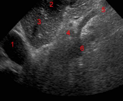
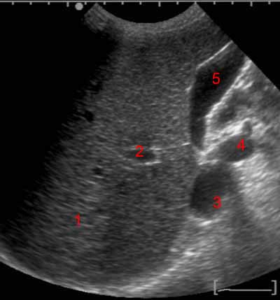
Abdominal ultrasound. Image 1. 1, Diaphragm. 2, Middle hepatic vein. 3, Left hepatic vein. 4, Inferior vena cava. 5, Right lobe of the liver. 6, Left lobe of the liver.
-
Abdominal ultrasound. Image 2. 1, Diaphragm. 2, Heart. 3, Inferior vena cava. 4, Left hepatic vein. 5, Left lobe of the liver.
-
Abdominal ultrasound. Image 3. 1, Heart. 2, Left lobe of the liver. 3, Left hepatic vein. 4, Celiac trunk. 5, Superior mesenteric artery. 6, Abdominal aorta.
-
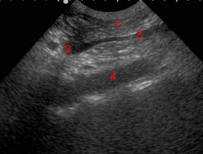
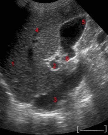
Abdominal ultrasound. Image 4. 1, Pancreas. 2, Portal confluence. 3, Superior mesenteric vein. 4, Abdominal aorta.
-
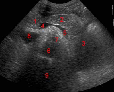
Abdominal ultrasound. Image 5. 1, Head of pancreas. 2, Body of pancreas. 3, Tail of pancreas. 4, Portal confluence. 5, Splenic vein. 6, Abdominal aorta. 7, Superior mesenteric artery. 8, Inferior vena cava. 9, Spine.
-
Abdominal ultrasound. Image 6. 1, Right lobe of the liver. 2, Portal vein. 3, Biliary tract. 4, Diaphragm. 5, Inferior vena cava. 6, Spine.
-
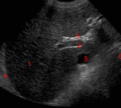
Abdominal ultrasound. Image 7. 1, Right lobe of the liver. 2, Branch of the portal vein. 3, Inferior vena cava. 4, Abdominal aorta. 5, Gallbladder.
-
Abdominal ultrasound. Image 8. 1, Right lobe of the liver. 2, Branch of the portal vein. 3, Inferior vena cava. 4, Hepatic vein. 5, Gallbladder. 6, infudibulum.
-
Abdominal ultrasound. Image 9. 1, Spleen. 2, Diaphragm.
-
Abdominal ultrasound. Image 10 of 10. 1, Renal cortex. 2, Pelvicalyceal system. 3, Renal sinus. 4, Liver.
