-
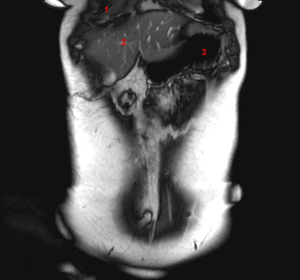
Image 1. Entero-MRI (anatomy), coronal view.1, Right lung. 2, Liver. 3, Stomach.
-
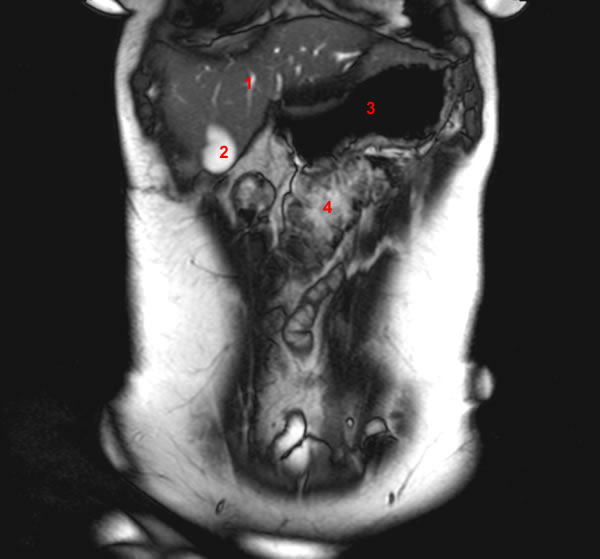
Image 2. Entero-MRI (anatomy), coronal view.1, Liver. 2, Gallbladder. 3, Stomach. 4, Transverse colon.
-
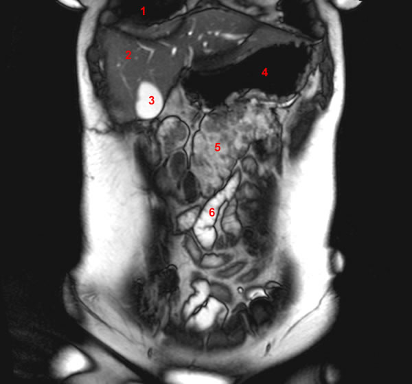
Image 3. Entero-MRI (anatomy), coronal view.1, Right lung. 2, Liver. 3, Gallbladder. 4, Stomach. 5, Transverse colon. 6, Small Intestine.
-
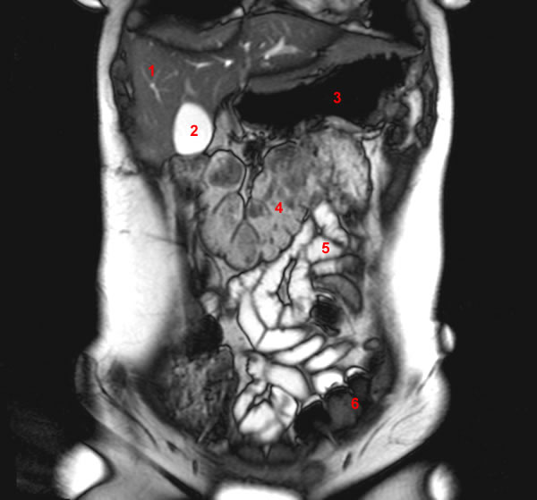
Image 4. Entero-MRI (anatomy), coronal view.1, Liver. 2, Gallbladder. 3, Stomach. 4, Transverse colon. 5, Small Intestine. 6, Sigmoid.
-
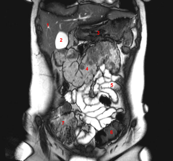
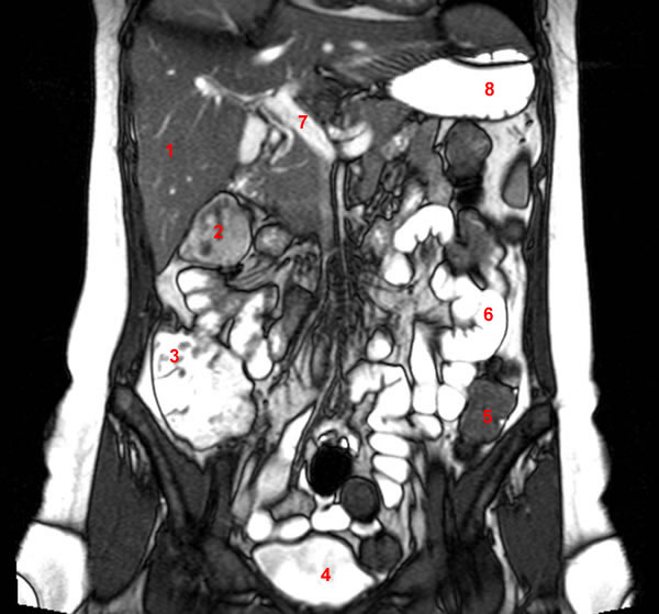
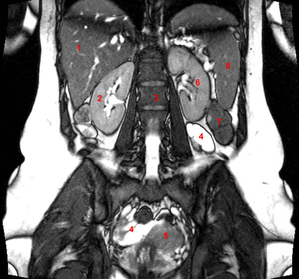
Image 5. Entero-MRI (anatomy), coronal view.1, Liver. 2, Gallbladder. 3, Stomach. 4, Transverse colon. 5, Small Intestine. 6, Sigmoid. 7, Cecum.
-
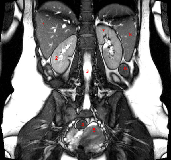
Image 6. Entero-MRI (anatomy), coronal view.1, Liver. 2, Gallbladder. 3, Transverse colon. 4, Stomach. 5, Splenic (or left colic) flexure. 6, Small Intestine. 7, Sigmoid. 8, Cecum.
-
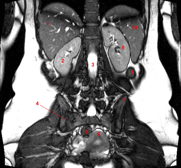
Image 7. Entero-MRI (anatomy), coronal view. 1, Liver. 2, Gallbladder. 3, Stomach. 4, Splenic (or left colic) flexure. 5, Transverse colon. 6, Small Intestine. 7, Iliac wing. 8, Cecum. 9, Sigmoid.
-
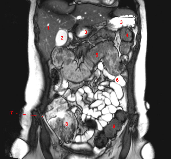
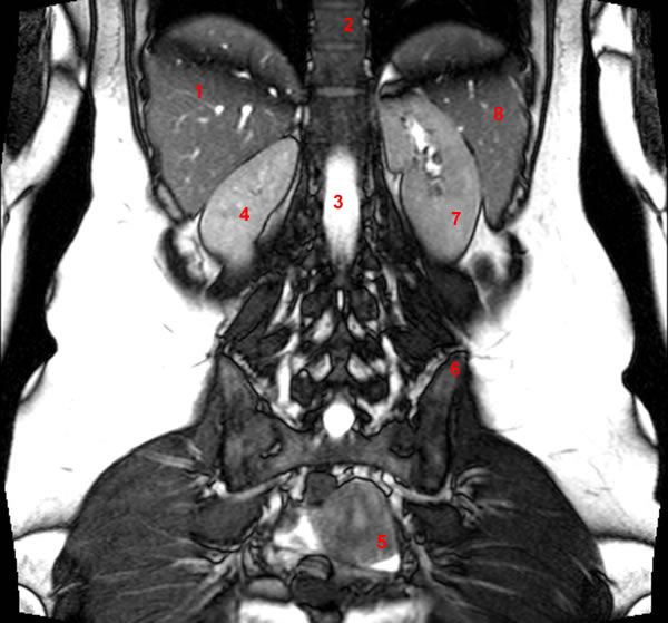
Image 8. Entero-MRI (anatomy), coronal view. 1, Liver. 2, Hepatic vein. 3, Hepatic (or the right colic) flexure. 4, Transverse colon. 5, Splenic (or left colic) flexure. 6, Stomach. 7, Small Intestine. 8, Cecum. 9, Sigmoid. 10, Iliac wing. 11, Bladder.
-
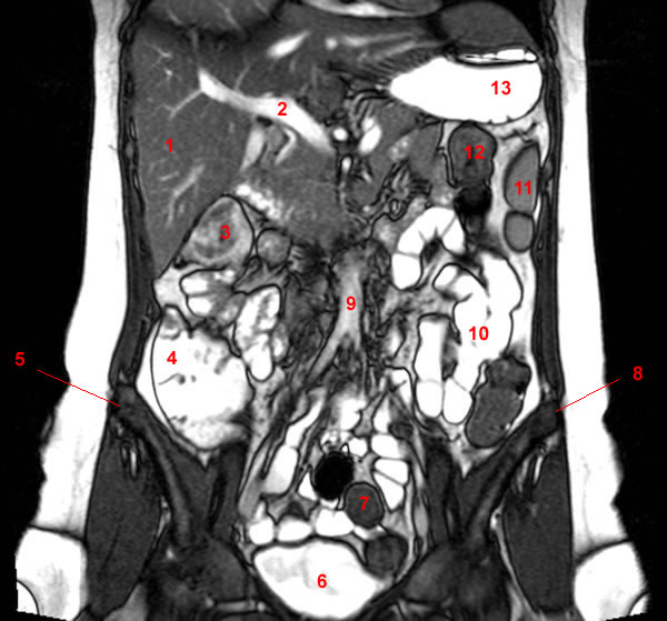
Image 9. Entero-MRI (anatomy), coronal view. 1, Hepatic vein. 2, Liver. 3, Hepatic (or the right colic) flexure. 4, Terminal ileum. 5, Cecum. 6, Stomach. 7, Splenic (or left colic) flexure. 8, Transverse colon. 9, Small Intestine. 10, Sigmoid. 11, Iliac wing. 12, Bladder.
-
Image 10. Entero-MRI (anatomy), coronal view. 1, Liver. 2, Gallbladder. 3, Hepatic (or the right colic) flexure. 4, Iliac wing. 5, Terminal ileum. 6, Cecum. 7, Bladder. 8, Sigmoid. 9, Small Intestine. 10, Transverse colon. 11, Splenic (or left colic) flexure. 12, Stomach.
-
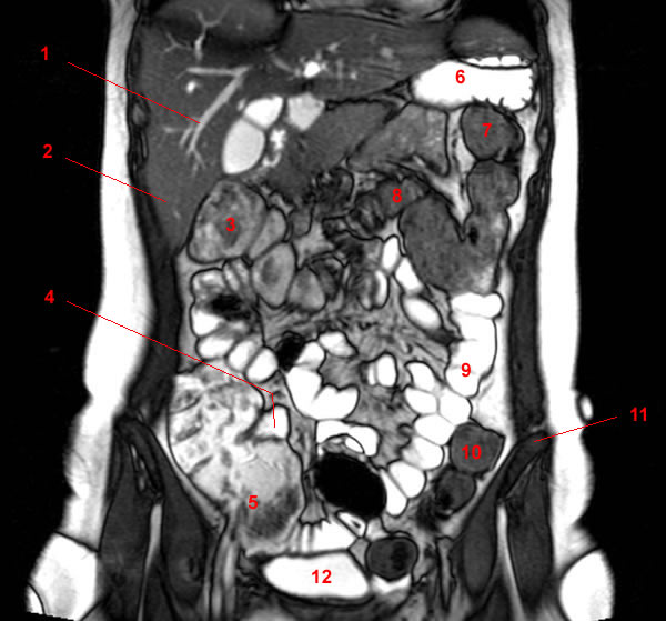
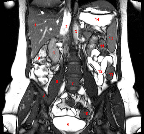
Image 11. Entero-MRI (anatomy), coronal view. 1, Liver. 2, Gallbladder. 3, Hepatic (or the right colic) flexure. 4, Iliac wing. 5, Cecum. 6, Bladder. 7, Sigmoid. 8, Small Intestine. 9, Superior mesenteric vein. 10, Splenic vein. 11, Descending colon. 12, Splenic (or left colic) flexure. 13, Stomach.
-
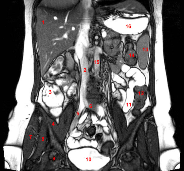
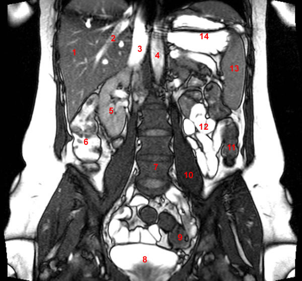
Image 12. Entero-MRI (anatomy), coronal view. 1, Liver. 2, Hepatic (or the right colic) flexure. 3, Ascending colon. 4, Bladder. 5, Iliac wing. 6, Sigmoid. 7, Superior mesenteric vein. 8, Splenic vein. 9, Portal vein. 10, Small Intestine. 11, Descending colon. 12, Splenic (or left colic) flexure. 13, Stomach.
-
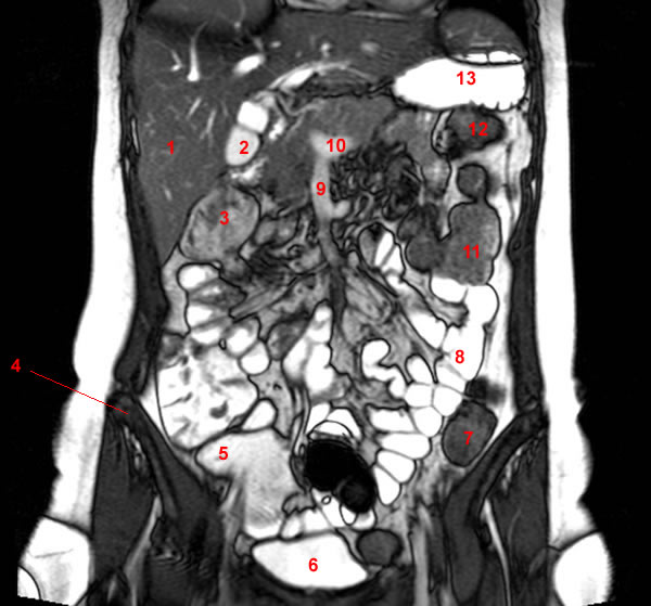
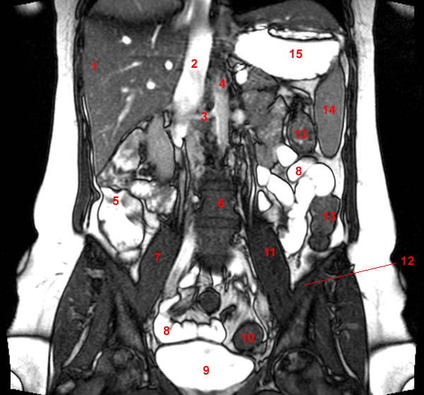
Image 13. Entero-MRI (anatomy), coronal view. 1, Liver. 2, Hepatic (or the right colic) flexure. 3, Ascending colon. 4, Bladder. 5, Sigmoid/Descending colon. 6, Small Intestine. 7, Portal vein. 8, Stomach.
-
Image 14. Entero-MRI (anatomy), coronal view. 1, Liver. 2, Portal vein. 3, Hepatic (or the right colic) flexure. 4, Ascending colon. 5, Iliac wing (right side). 6, Bladder. 7, Sigmoid. 8, Iliac wing (left side). 9, Abdominal aorta. 10, Small Intestine. 11, Spleen. 12, Colon. 13, Stomach.
-
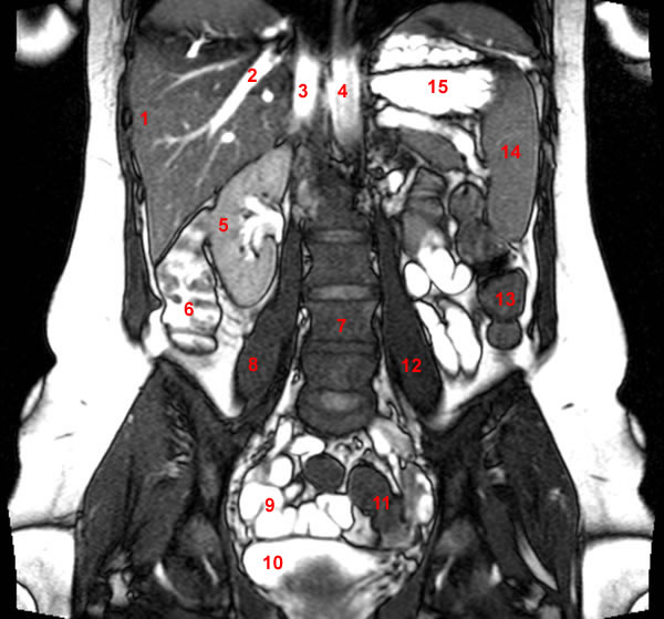
Image 15. Entero-MRI (anatomy), coronal view. 1, Liver. 2, Portal vein. 3, Hepatic (or the right colic) flexure. 4, Ascending colon. 5, Inferior vena cava. 6, Aorta 7, Right common iliac artery. 8, Left common iliac artery. 9, Small Intestine. 10, Bladder. 11, Sigmoid 12, Descending colon/Sigmoid. 13, Descending colon. 14, Spleen. 15, Stomach.
-
Image 16. Entero-MRI (anatomy), coronal view. 1, Liver. 2, Ascending colon. 3, Inferior vena cava. 4, Left iliac vein. 5, Right psoas muscle. 6, Iliac wing (right side). 7, Right femoral head. 8, Bladder. 9, Sigmoid. 10, Small Intestine. 11, Descending colon. 12, Spleen. 13, Descending colon. 14, Stomach.
-
Image 17. Entero-MRI (anatomy), coronal view. 1, Liver. 2, Inferior vena cava. 3, Ascending colon. 4, Iliacus muscle. 5, Right iliac vein. 6, Left iliac vein. 7, Gluteus medius muscle. 8, Gluteus minimus muscle. 9, Femoral head. 10, Bladder. 11, Small Intestine. 12, Descending colon. 13, Spleen. 14, Descending colon. 15, Abdominal aorta. 16, Stomach.
-
Image 18. Entero-MRI (anatomy), coronal view. 1, Liver. 2, Inferior vena cava. 3, Right renal artery. 4, Abdominal aorta. 5, Ascending colon. 6, Lumbar spine. 7, Right psoas muscle. 8, Small Intestine. 9, Bladder. 10, Sigmoid. 11, Left psoas muscle. 12, Iliacus muscle. 13, Descending colon. 14, Spleen. 15, Stomach.
-
Image 19. Entero-MRI (anatomy), coronal view. 1, Liver. 2, Inferior vena cava. 3, Abdominal aorta. 4, Right kidney. 5, Ascending colon. 6, Right psoas muscle. 7, Lumbar spine. 8, Iliacus muscle. 9, Bladder. 10, Sigmoid. 11, Descending colon. 12, Small Intestine. 13, Spleen. 14, Stomach.
-
Image 20. Entero-MRI (anatomy), coronal view. 1, Liver. 2, Hepatic vein. 3, Inferior vena cava. 4, Abdominal aorta. 5, Right kidney. 6, Ascending colon. 7, Lumbar spine. 8, Bladder. 9, Sigmoid. 10, Left psoas muscle. 11, Descending colon. 12, Small Intestine. 13, Spleen. 14, Stomach.
-
Image 21. Entero-MRI (anatomy), coronal view. 1, Liver. 2, Hepatic vein. 3, Inferior vena cava. 4, Abdominal aorta. 5, Right kidney. 6, Ascending colon. 7, Lumbar spine. 8, Right psoas muscle. 9, Small Intestine. 10, Bladder. 11, Sigmoid. 12, Left psoas muscle. 13, Descending colon. 14, Spleen. 15, Stomach.
-
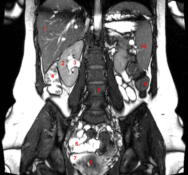
Image 22. Entero-MRI (anatomy), coronal view. 1, Liver. 2, Right kidney. 3, Right renal pelvis. 4, Ascending colon. 5, Lumbar spine. 6, Small Intestine. 7, Bladder. 8, Uterus. 9, Descending colon. 10, Spleen.
-
Image 23. Entero-MRI (anatomy), coronal view. 1, Liver. 2, Right kidney. 3, Right renal pelvis. 4, Ascending colon. 5, Right psoas muscle. 6, Lumbar spine. 7, Small Intestine. 8, Uterus. 9, Left psoas muscle. 10, Descending colon. 11, Left kidney. 12, Spleen.
-
Image 24. Entero-MRI (anatomy), coronal view. 1, Liver. 2, Right kidney. 3, Lumbar spine. 4, Small Intestine. 5, Uterus. 6, Left kidney. 7, Descending colon. 8, Spleen.
-
Image 25. Entero-MRI (anatomy), coronal view. 1, Liver. 2, Right kidney. 3, Ascending colon. 4, Uterus. 5, Vertebral canal. 6, Small Intestine. 7, Descending colon. 8, Left kidney. 9, Spleen.
-
Image 26. Entero-MRI (anatomy), coronal view. 1, Liver. 2, Right kidney. 3, Vertebral canal. 4, Rectum. 5, Uterus. 6, Descending colon. 7, Left kidney. 8, Spleen.
-
Image 27. Entero-MRI (anatomy), coronal view. 1, Liver. 2, Right kidney. 3, Vertebral canal. 4, Sacroiliac joint (right side). 5, Rectum. 6, Uterus. 7, Iliac wing (left side). 8, Descending colon. 9, Left kidney. 10, Spleen.
-
Image 28 of 28. Entero-MRI (anatomy), coronal view. 1, Liver. 2, Spine. 3, Vertebral canal. 4, Right kidney. 5, Uterus. 6, Iliac wing (left side). 7, Left kidney. 8, Spleen.