-
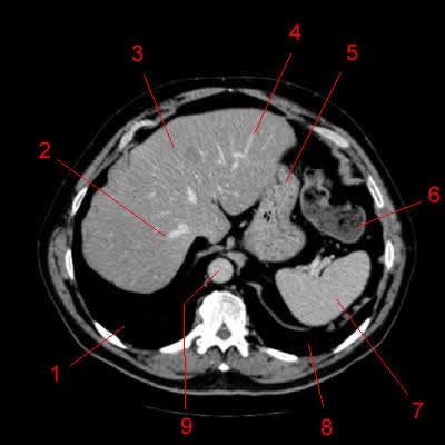
Image 1. Atlas of CT Anatomy of the Abdomen. Axial reconstruction.
1, Right lung. 2, Right hepatic vein. 3, Liver. 4, Left hepatic vein. 5, Stomach. 6, Left colic flexure (splenic flexure of the colon). 7, Spleen. 8, Left lung. 9, Aorta.
-
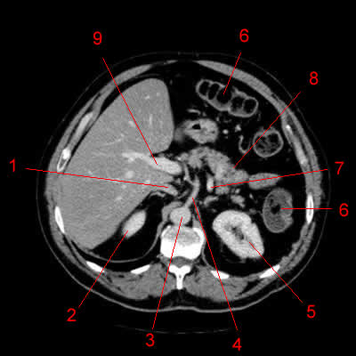
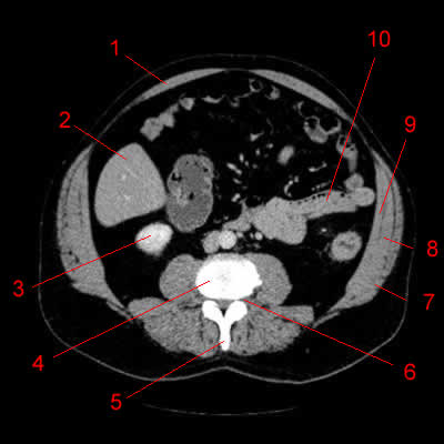
Image 2. Atlas of CT Anatomy of the Abdomen. Axial reconstruction.
1, Right lung. 2, Aorta. 3, Left lung. 4, Left adrenal. 5, Spleen. 6, Splenic artery. 7, Colon. 8, Portal vein. 9, Hepatic vein. 10, Liver.
-
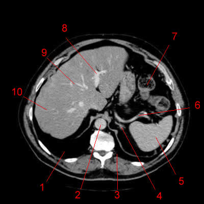
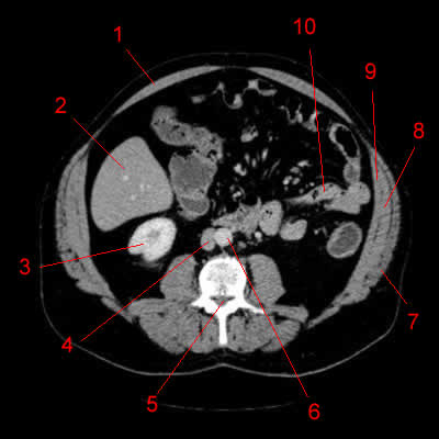
Image 3. Atlas of CT Anatomy of the Abdomen. Axial reconstruction.
1, Diaphragm. 2, Aorta. 3, Left adrenal. 4, Top of the left kidney. 5, Spleen. 6, Splenic artery. 7, Colon. 8, Stomach. 9, Portal vein. 10, Liver. 11, Rib.
-
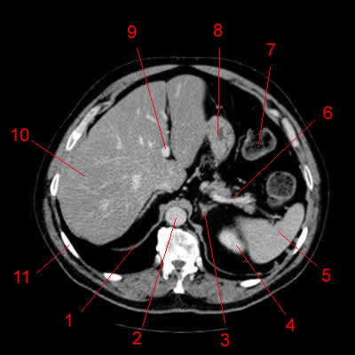
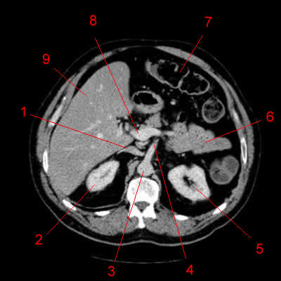
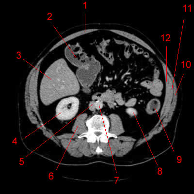
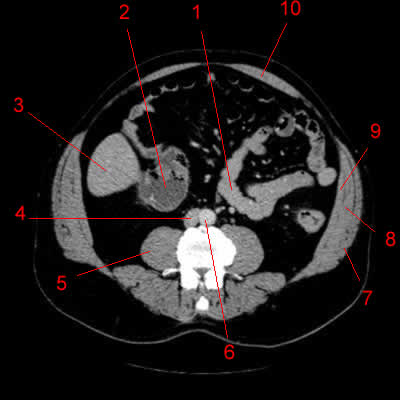
Image 4. Atlas of CT Anatomy of the Abdomen. Axial reconstruction.
1, Inferior vena cava. 2, Liver. 3, Right adrenal. 4, Diaphragmatic crus. 5, Abdominal aorta. 6, Left adrenal. 7, Left kidney. 8, Spleen. 9, Pancreas. 10, Colon. 11, Portal vein.
-
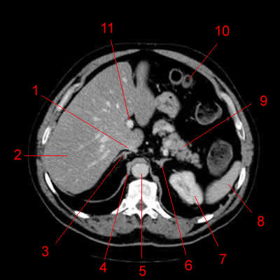
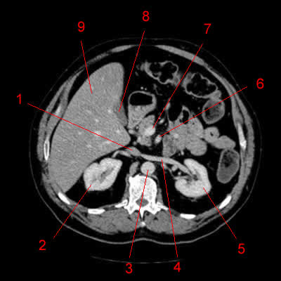
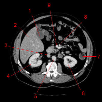
Image 5. Atlas of CT Anatomy of the Abdomen. Axial reconstruction.
1, Inferior vena cava. 2, Top of the right kidney. 3, Abdominal aorta. 4, Celiac truncus. 5, Left kidney. 6, Colon. 7, Splenic vein. 8, Pancreas. 9, Portal vein.
-
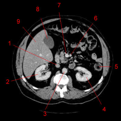
Image 6. Atlas of CT Anatomy of the Abdomen. Axial reconstruction.
1, Inferior vena cava. 2, Right kidney. 3, Abdominal aorta. 4, Superior mesenteric artery. 5, Left kidney. 6, Small bowel. 7, Colon. 8, Portal vein. 9, Liver.
-
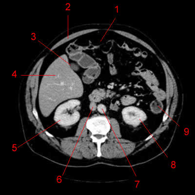
Image 7. Atlas of CT Anatomy of the Abdomen. Axial reconstruction.
1, Inferior vena cava. 2, Right kidney. 3, Aorta. 4, Left renal vein. 5, Left kidney. 6, Superior mesenteric artery. 7, Superior mesenteric vein. 8, Gallbladder. 9, Liver.
-
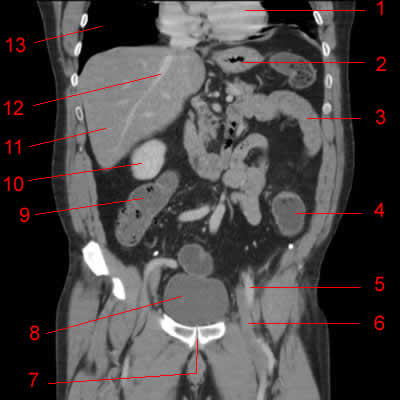
Image 8. Atlas of CT Anatomy of the Abdomen. Axial reconstruction.
1, Inferior vena cava. 2, Right kidney. 3, Origin of the right renal artery. 4, Aorta. 5, Left kidney. 6, Left colon. 7, Superior mesenteric artery. 8, Superior mesenteric vein. 9, Gallbladder. 10, Liver.
-
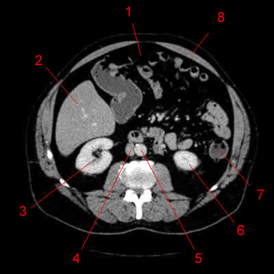
Image 9. Atlas of CT Anatomy of the Abdomen. Axial reconstruction.
1, Inferior vena cava. 2, Right kidney. 3, Aorta. 4, Left kidney. 5, Left colon. 6, Superior mesenteric artery. 7, Superior mesenteric vein. 8, Gallbladder. 9, Liver.
-
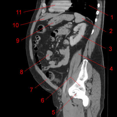
Image 10. Atlas of CT Anatomy of the Abdomen. Axial reconstruction.
1, Gallbladder. 2, Liver. 3, Inferior vena cava. 4, Right kidney. 5, Aorta. 6, Left kidney. 7, Left colon. 8, Superior mesenteric artery. 9, Superior mesenteric vein.
-
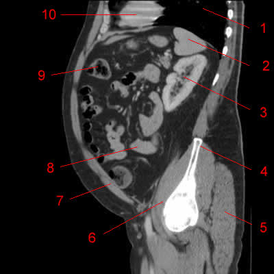
Image 11. Atlas of CT Anatomy of the Abdomen. Axial reconstruction.
1, Transverse colon. 2, Rectus abdominis muscle. 3, Gallbladder. 4, Liver. 5, Right kidney. 6, Inferior vena cava. 7, Aorta. 8, Left kidney. 9, Left colon.
-
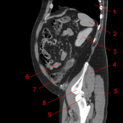
Image 12. Atlas of CT Anatomy of the Abdomen. Axial reconstruction.
1, Transverse colon. 2, Liver. 3, Right kidney. 4, Inferior vena cava. 5, Aorta. 6, Lower pole of the left kidney. 7, Left colon. 8, Rectus abdominis muscle.
-
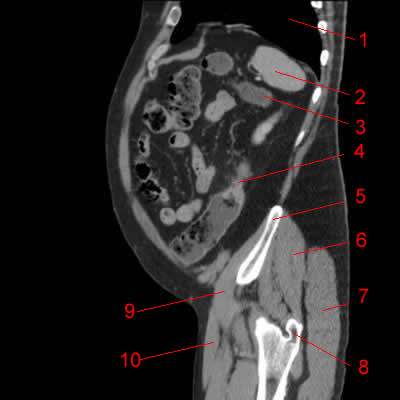
Image 13. Atlas of CT Anatomy of the Abdomen. Axial reconstruction.
1, Rectus abdominis muscle. 2, Colon. 3, Liver. 4, Right kidney. 5, Inferior vena cava. 6, Psoas muscle. 7, Aorta. 8, Lower pole of the left kidney. 9, Left colon. 10, External oblique muscle. 11, Internal oblique muscle. 12, Transversus abdominis muscle.
-
Image 14. Atlas of CT Anatomy of the Abdomen. Axial reconstruction.
1, Rectus abdominis muscle. 2, Liver. 3, Right kidney. 4, Inferior vena cava. 5, Vertebral canal. 6, Aorta. 7, External oblique muscle. 8, Internal oblique muscle. 9, Transversus abdominis muscle. 10, Small intestine.
-
Image 15. Atlas of CT Anatomy of the Abdomen. Axial reconstruction.
1, Rectus abdominis muscle. 2, Liver. 3, Inferior pole of the right kidney. 4, Vertebral body. 5, Spinous process. 6, Intervertebral foramen. 7, External oblique muscle. 8, Internal oblique muscle. 9, Transversus abdominis muscle. 10, Small intestine.
-
Image 16. Atlas of CT Anatomy of the Abdomen. Axial reconstruction.
1, Small intestine. 2, Colon. 3, Liver. 4, Inferior vena cava. 5, Psoas muscle. 6, Aorta. 7, External oblique muscle. 8, Internal oblique muscle. 9, Transversus abdominis muscle. 10, Rectus abdominis muscle.
-
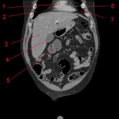
Image 17. Atlas of CT Anatomy of the Abdomen. Coronal reconstruction.
1, Heart. 2, Right lung. 3, Liver. 4, Gallbladder. 5, Colon. 6, Rib. 7, Left lung.
-
Image 18. Atlas of CT Anatomy of the Abdomen. Coronal reconstruction.
1, Right lung. 2, Liver. 3, Gallbladder. 4, Caecum. 5, Bladder. 6, Heart 7, Stomach. 8, Colon. 9, Small bowel. 10, Sigmoid colon.
-
Image 19. Atlas of CT Anatomy of the Abdomen. Coronal reconstruction.
1, Right lung. 2, Portal vein. 3, Liver. 4, Caecum. 5, Bladder. 6, Sigmoid colon. 7, Small bowel. 8, Colon. 9, Heart.
-
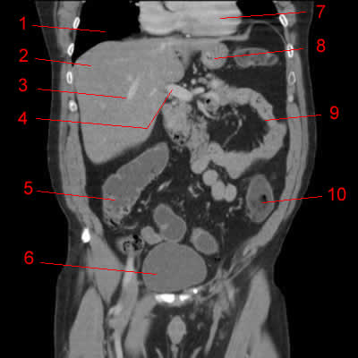
Image 20. Atlas of CT Anatomy of the Abdomen. Coronal reconstruction.
1, Right lung. 2, Portal vein. 3, Liver. 4, Right colon. 5, Bladder. 6, Sigmoid colon. 7, Small intestine. 8, Superior mesenteric vein. 9, Heart.
-
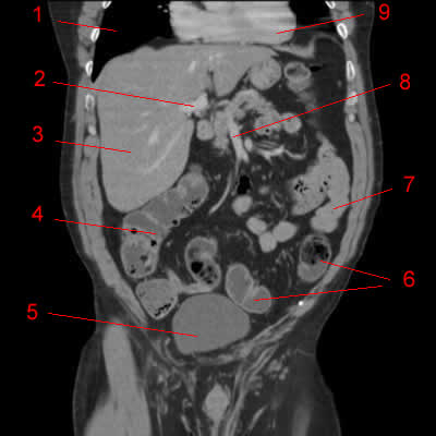
Image 21. Atlas of CT Anatomy of the Abdomen. Coronal reconstruction.
1, Right lung. 2, Liver. 3, Hepatic vein. 4, Portal vein. 5, Right colon. 6, Bladder. 7, Heart. 8, Stomach. 9, Small intestine. 10, Left colon.
-
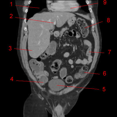
Image 22. Atlas of CT Anatomy of the Abdomen. Coronal reconstruction.
1, Heart. 2, Stomach. 3, Small bowel. 4, Left colon. 5, Left superficial femoral artery. 6, Left superficial femoral vein. 7, Pubic symphysis. 8, Bladder. 9, Right colon 10, Right kidney. 11, Liver. 12, Hepatic vein. 13, Right lung.
-
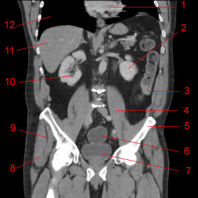
Image 23. Atlas of CT Anatomy of the Abdomen. Coronal reconstruction.
1, Left lung. 2, Colic tumor. 3, External oblique muscle. 4, Internal oblique muscle. 5, Transversus abdominis muscle. 6, Bladder. 7, Pubic symphysis. 8, Aorta. 9, Inferior vena cava. 10, Right kidney. 11, Liver. 12, Heart.
-
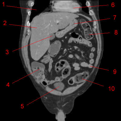
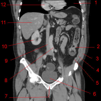
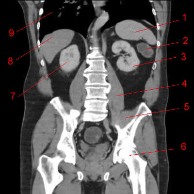
Image 24. Atlas of CT Anatomy of the Abdomen. Coronal reconstruction.
1, Heart. 2, Left kidney. 3, Colic tumor. 4, Psoas muscle. 5, Iliac wing. 6, Rectum. 7, Bladder. 8, Gluteus medius muscle. 9, Gluteus minimus muscle. 10, Right kidney. 11, Liver. 12, Right lung.
-
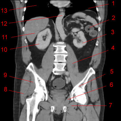
Image 25. Atlas of CT Anatomy of the Abdomen. Coronal reconstruction.
1, Left lung. 2, Left colon. 3, Left kidney. 4, Psoas muscle. 5, Acetabulum. 6, Obturator internus muscle. 7, Obturator externus muscle. 8, Gluteus medius muscle. 9, Gluteus minimus muscle. 10, Right kidney. 11, Liver. 12, Aorta. 13, Right lung.
-
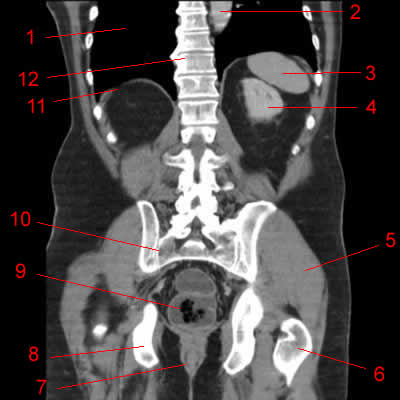
Image 26. Atlas of CT Anatomy of the Abdomen. Coronal reconstruction.
1, Spleen. 2, Left colic flexure (splenic flexure of the colon). 3, Left kidney. 4, Psoas muscle. 5, Iliac muscle. 6, Femoral head. 7, Right kidney. 8, Liver. 9, Right lung.
-
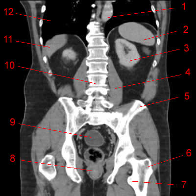
Image 27. Atlas of CT Anatomy of the Abdomen. Coronal reconstruction.
1, Aorta. 2, Spleen. 3, Left kidney. 4, Psoas muscle. 5, Ilium. 6, Greater trochanter. 7, Lesser trochanter. 8, Anal canal 9, Rectum. 10, Lumbar spine. 11, Liver. 12, Right lung.
-
Image 28. Atlas of CT Anatomy of the Abdomen. Coronal reconstruction.
1, Right lung. 2, Aorta. 3, Spleen. 4, Left kidney. 5, Gluteus maximus muscle. 6, Greater trochanter. 7, Anal canal. 8, Ischium. 9, Rectum. 10, Sacroiliac joint. 11, Diaphragm. 12, Thoracic spine.
-
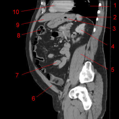
Image 29. Atlas of CT Anatomy of the Abdomen. Sagittal reconstruction.1, Left lung. 2, Spleen. 3, Colon. 4, Colic tumor. 5, Iliac wing. 6, Gluteus medius muscle. 7, Gluteus minimus muscle. 8, Gluteus maximus muscle. 9, Sartorius muscle. 10, Left femur.
-
Image 30. Atlas of CT Anatomy of the Abdomen. Sagittal reconstruction.
1, Left lung. 2, Spleen. 3, Colon. 4, Colic tumor. 5, Iliac wing. 6, Gluteus medius muscle. 7, Gluteus maximus muscle. 8, Greater trochanter. 9, Iliac muscle. 10, Sartorius muscle.
-
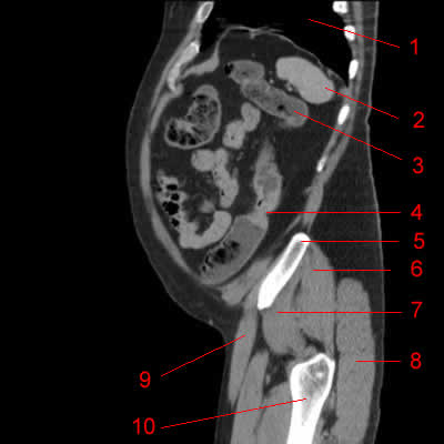
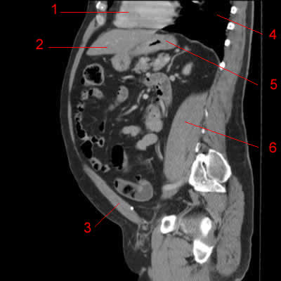
Image 31. Atlas of CT Anatomy of the Abdomen. Sagittal reconstruction.
1, Left lung. 2, Spleen. 3, Left kidney. 4, Gluteus medius muscle. 5, Gluteus maximus muscle. 6, Small bowel. 7, Colon. 8, Acetabulum. 9, Femoral head.
-
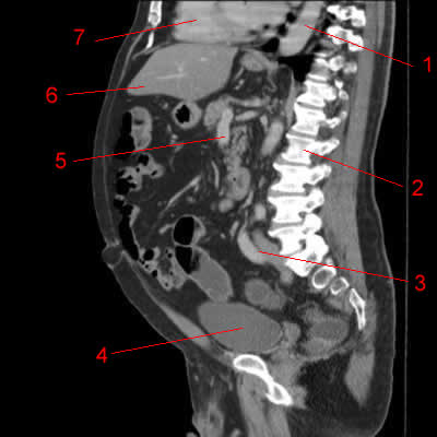
Image 32. Atlas of CT Anatomy of the Abdomen. Sagittal reconstruction.
1, Left lung. 2, Spleen. 3, Left kidney. 4, Iliac wing. 5, Gluteus medius muscle. 6, Gluteus maximus muscle. 7, Femoral head. 8, Heart. 9, Small bowel. 10, Colon. 11, Iliac muscle. 12, Sartorius muscle.
-
Image 33. Atlas of CT Anatomy of the Abdomen. Sagittal reconstruction.
1, Left lung. 2, Spleen. 3, Left kidney. 4, Iliac wing. 5, Gluteus maximus muscle. 6, Iliac muscle. 7, Rectus abdominis muscle. 8, Small bowel. 9, Colon. 10, Heart.
-
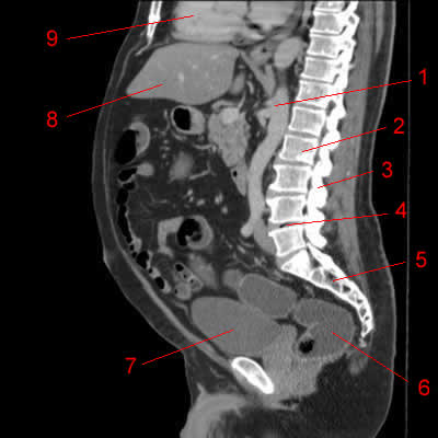
Image 34. Atlas of CT Anatomy of the Abdomen. Sagittal reconstruction.
1, Left lung. 2, Spleen. 3, Left kidney. 4, Psoas muscle. 5, Femoral head. 6, Acetabulum. 7, Colon. 8, Small bowel. 9, Stomach. 10, Liver. 11, Heart.
-
Image 35. Atlas of CT Anatomy of the Abdomen. Sagittal reconstruction.
1, Left lung. 2, Spleen. 3, Stomach. 4, Left kidney. 5, Psoas muscle. 6, Rectus abdominis muscle. 7, Small bowel. 8, Colon. 9, Liver. 10, Heart.
-
Image 36. Atlas of CT Anatomy of the Abdomen. Sagittal reconstruction.
1, Heart. 2, Liver. 3, Rectus abdominis muscle. 4, Left lung. 5, Stomach. 6, Psoas muscle.
-
Image 37. Atlas of CT Anatomy of the Abdomen. Sagittal reconstruction.
1, Aorta. 2, Psoas muscle. 3, Iliac artery. 4, Bladder. 5, Liver. 6, Heart.
-
Image 38. Atlas of CT Anatomy of the Abdomen. Sagittal reconstruction.
1, Aorta. 2, Lumbar spine. 3, Iliac vessels. 4, Bladder. 5, Superior mesenteric vein. 6, Liver. 7, Heart.
-
Image 39. Atlas of CT Anatomy of the Abdomen. Sagittal reconstruction.
1, Aorta. 2, Vertebral body. 3, Spinous process. 4, Intervertebral disc. 5, Sacrum. 6, Rectum. 7, Bladder. 8, Liver. 9, Heart.
-
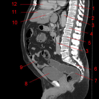
Image 40 of 40. Atlas of CT Anatomy of the Abdomen. Sagittal reconstruction.
1, Vertebral body (Thoracic spine). 2, Spinous process. 3, Aorta. 4, Vertebral body (lumbar spine, L1). 5, Intervertebral disc. 6, Sacrum. 7, Rectum. 8, Bladder. 9, Small bowel. 10, Hepatic vein. 11, Liver. 12, Heart.