-
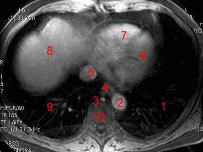
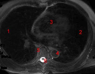
Atlas of MRI Anatomy of the Abdomen. Axial T1-weighted image. Image 1.
1, Lower lobe of the left lung. 2, Thoracic aorta. 3, Azygos vein. 4, Esophagus. 5, Heart. 6, Dome of the liver. 7, Lower lobe of the right lung.
-
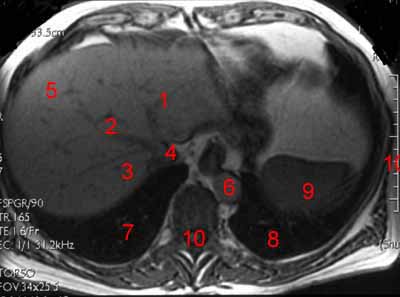
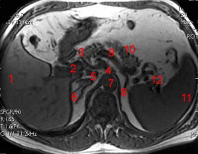
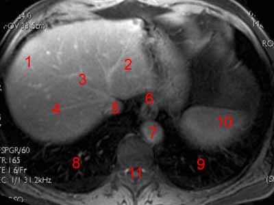
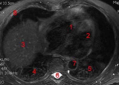
Atlas of MRI Anatomy of the Abdomen. Axial T1-weighted image. Image 2.
1, Left hepatic vein. 2, Middle hepatic vein. 3, Right hepatic vein. 4, Inferior vena cava. 5, Dome of the liver. 6, Abdominal aorta. 7, Lower lobe of the right lung. 8, Inferior lobe of the left lung. 9, Spleen. 10, Vertebral canal. 11, Rib.
-
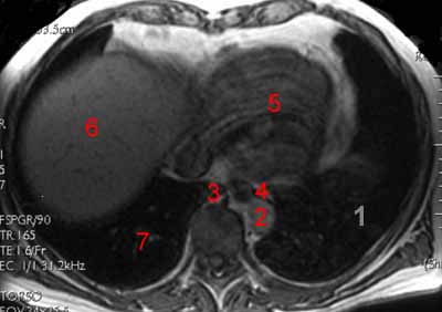
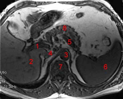
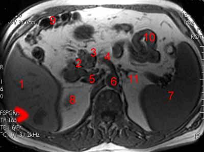
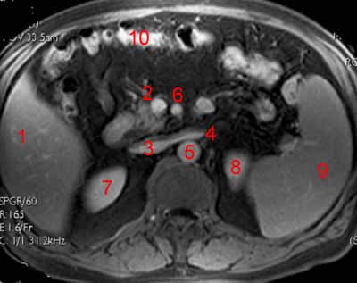
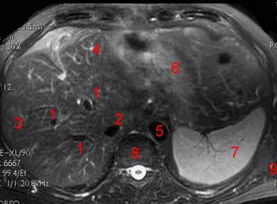
Atlas of MRI Anatomy of the Abdomen. Axial T1-weighted image. Image 3.
1, Porta hepatis. 2, Portal vein. 3, Stomach. 4, Diaphragm. 5, Right lobe of the liver. 6, Left lobe of the liver. 7, Spleen. 8, Lung. 9, Abdominal aorta.
-
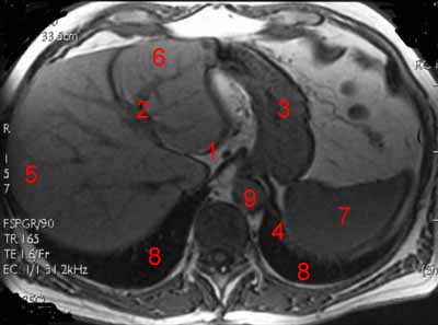
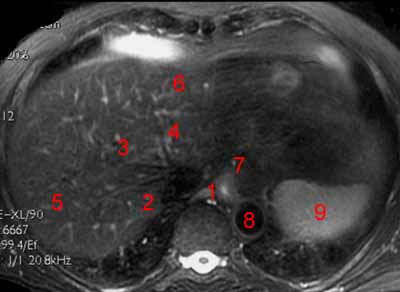
Atlas of MRI Anatomy of the Abdomen. Axial T1-weighted image. Image 4.
1, Crus of diaphragm. 2, Right lung. 3, Left lobe of the liver. 4, Portal vein. 5, Right lobe of the liver. 6, Stomach. 7, Pancreas. 8, Aorta. 9, Spleen. 10, Longuissimus dorsi. 11, Vertebral canal.
-
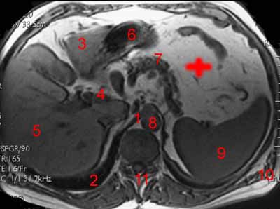
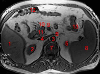
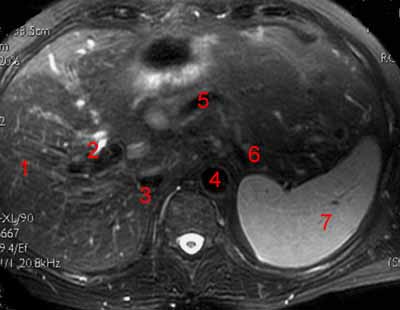
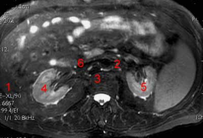
Atlas of MRI Anatomy of the Abdomen. Axial T1-weighted image. Image 5.
1, Portal vein. 2, Right branch of the portal vein. 3, Aorta. 4, Inferior vena cava. 5, Splenic vein. 6, Spleen. 7, Vertebral body. 8, Body of the pancreas.
-
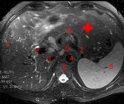
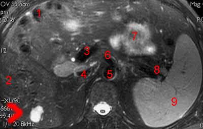
Atlas of MRI Anatomy of the Abdomen. Axial T1-weighted image. Image 6.
1, Liver. 2, Portal vein. 3, Hepatic artery. 4, Celiac trunk. 5, Inferior vena cava. 6, Right adrenal. 7, Aorta. 8, Left adrenal. 9, Splenic vein. 10, Pancreas. 11, Spleen. 12, Splenic hilus.
-
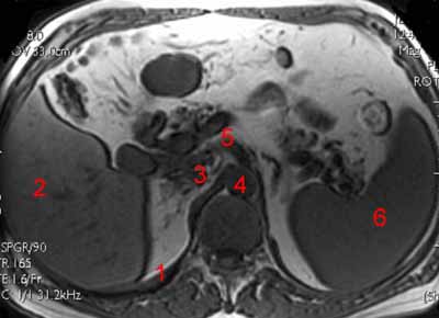
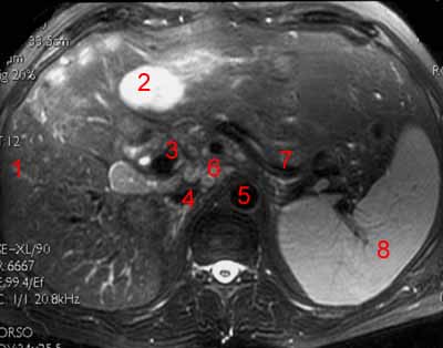
Atlas of MRI Anatomy of the Abdomen. Axial T1-weighted image. Image 7.
1, Diaphragm. 2, Liver. 3, Inferior vena cava. 4, Aorta. 5, Celiac trunk. 6, Spleen.
-
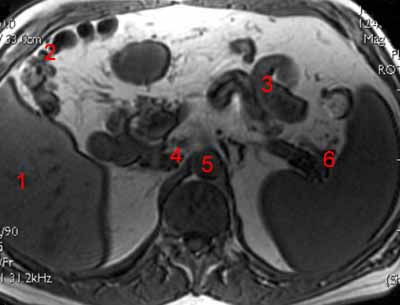
Atlas of MRI Anatomy of the Abdomen. Axial T1-weighted image. Image 8.
1, Liver. 2, Colon. 3, Small bowel. 4, Inferior vena cava. 5, Aorta. 6, Splenic hilus.
-
Atlas of MRI Anatomy of the Abdomen. Axial T1-weighted image. Image 9.
1, Liver. 2, Pancreas. 3, Superior mesenteric vein. 4, Superior mesenteric artery. 5, Inferior vena cava. 6, Aorta. 7, Spleen. 8, Top of the right kidney. 9, Transverse colon. 10, Small bowel. 11, Left adrenal.
-
Atlas of MRI Anatomy of the Abdomen. Axial T1-weighted image. Image 10.
1, Liver. 2, Right kidney. 3, Aorta. 4, Inferior vena cava. 5, Left kidney. 6, Left renal vein. 7, Spine. 8, Spleen. 9, Superior mesenteric artery. 10, Superior mesenteric vein. 11, Pancreas. 12, Colon. 13, Small bowel.
-
Atlas of MRI Anatomy of the Abdomen. Axial T1-weighted image after gadolinium. Image 11.
1, Left lung. 2, Thoracic aorta. 3, Azygos vein. 4, Esophagus. 5, Inferior vena cava. 6, Left ventricle. 7, Right ventricle. 8, Dome of the liver. 9, Right lung. 10, Vertebral body.
-
Atlas of MRI Anatomy of the Abdomen. Axial T1-weighted image after gadolinium. Image 12.
1, Liver. 2, Left hepatic vein. 3, Middle hepatic vein. 4, Right hepatic vein. 5, Inferior vena cava. 6, Esophagus. 7, Abdominal aorta. 8, Right inferior pulmonary lobe. 9, Left inferior pulmonary lobe. 10, Spleen. 11, Vertebral canal.
-
Atlas of MRI Anatomy of the Abdomen. Axial T1-weighted image after gadolinium. Image 13.
1, Left lobe of the liver. 2, Portal vein. 3, Ligamentum venosum. 4, Right lobe of the liver. 5, Abdominal aorta. 6, Stomach. 7, Crus of diaphragm. 8, Spleen. 9, Right lung. 10, Left lung.
-
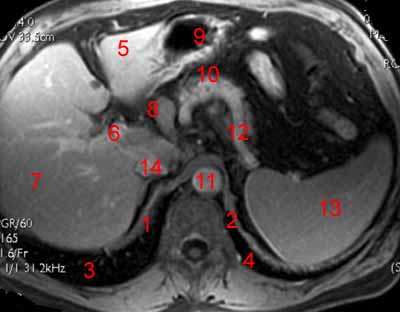
Atlas of MRI Anatomy of the Abdomen. Axial T1-weighted image after gadolinium. Image 14.
1, Right diaphragm. 2, Left diaphragm. 3, Right lung. 4, Left lung. 5, Left lobe of the liver. 6, Portal vein. 7, Right lobe of the liver. 8, Porta hepatis. 9, Stomach. 10, Pancreas. 11, Aorta. 12, Splenic artery. 13, Spleen. 14, Inferior vena cava.
-
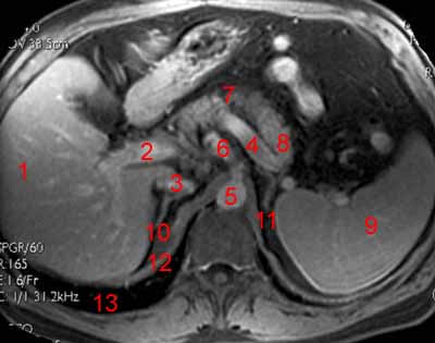
Atlas of MRI Anatomy of the Abdomen. Axial T1-weighted image after gadolinium. Image 15.
1, Liver. 2, Portal vein. 3, Inferior vena cava. 4, Splenic vein. 5, Aorta. 6, Celiac trunk. 7, Body of the pancreas.. 8, Pancratic tail. 9, Spleen. 10, Right adrenal. 11, Left adrenal. 12, Diaphragm. 13, Lung.
-
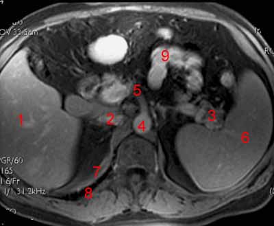
Atlas of MRI Anatomy of the Abdomen. Axial T1-weighted image after gadolinium. Image 16.
1, Liver. 2, Inferior vena cava. 3, Splenic vessels. 4, Aorta. 5, Superior mesenteric artery 6, Spleen. 7, Diaphragm. 8, Lung. 9, Small bowel.
-
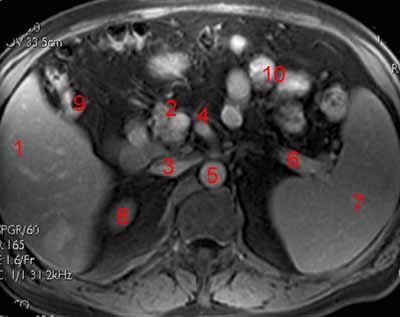
Atlas of MRI Anatomy of the Abdomen. Axial T1-weighted image after gadolinium. Image 17.
1, Liver. 2, Superior mesenteric vein. 3, Inferior vena cava. 4, Superior mesenteric artery. 5, Abdominal aorta. 6, Splenic vein. 7, Spleen. 8, Top of the right kidney. 9, Hepatic flexure of the colon. 10 Small bowel.
-
Atlas of MRI Anatomy of the Abdomen. Axial T1-weighted image after gadolinium. Image 18.
1, Liver. 2, Superior mesenteric vein. 3, Inferior vena cava. 4, Left renal vein. 5, Abdominal aorta. 6, Superior mesenteric artery. 7, Right kidney. 8, Left kidney. 9, Spleen. 10, Colon.
-
Atlas of MRI Anatomy of the Abdomen. Axial T2-weighted fat-suppressed image. Image 19.
1, Right lung. 2, Left lung. 3, Heart. 4, Aorta. 5, Vertebral body. 6, Spinal cord.
-
Atlas of MRI Anatomy of the Abdomen. Axial T2-weighted fat-suppressed image. Image 20.
1, Right ventricle. 2, Left ventricle. 3, Dome of the liver. 4, Inferior lobe of the rightt lung. 5, Inferior lobe of the left lung. 6, Vertebral canal. 7, Descending aorta. 8, Rib.
-
Atlas of MRI Anatomy of the Abdomen. Axial T2-weighted fat-suppressed image. Image 21.
1, Crus of diaphragm. 2, Right hepatic vein. 3, Middle hepatic vein. 4, Left hepatic vein. 5, Right lobe of the liver. 6, Left lobe of the liver. 7, Esophagus. 8, Aorta. 9, Spleen.
-
Atlas of MRI Anatomy of the Abdomen. Axial T2-weighted fat-suppressed image. Image 22.
1, Hepatic vein. 2, Inferior vena cava. 3, Right lobe of the liver. 4, Left lobe of the liver. 5, Aorta. 6, Stomach. 7, Spleen. 8, Vertebral body. 9, Longuissimus dorsi.
-
Atlas of MRI Anatomy of the Abdomen. Axial T2-weighted fat-suppressed image. Image 23.
1, Liver. 2, Portal vein. 3, Inferior vena cava. 4, Aorta. 5, Splenic artery. 6, Splenic vein. 7, Spleen.
-
Atlas of MRI Anatomy of the Abdomen. Axial T2-weighted fat-suppressed image. Image 24.
1, Liver. 2, Portal vein. 3, Inferior vena cava. 4, Aorta. 5, Origin of the celiac trunk. 6, Splenic artery. 7, Splenic vein. 8, Spleen. 9, Vertebral canal.
-
Atlas of MRI Anatomy of the Abdomen. Axial T2-weighted fat-suppressed image. Image 25.
1, Liver. 2, Gallbladder. 3, Portal confluence. 4, Inferior vena cava. 5, Aorta. 6, Superior mesenteric artery. 7, Splenic vein. 8, Spleen.
-
Atlas of MRI Anatomy of the Abdomen. Axial T2-weighted fat-suppressed image. Image 26.
1, colon. 2, Liver. 3, Portal confluence. 4, Inferior vena cava. 5, Aorta. 6, Superior mesenteric artery. 7, Small bowel. 8, Splenic vein. 9, Spleen.
-
Atlas of MRI Anatomy of the Abdomen. Axial T2-weighted fat-suppressed image. Image 27 of 27.
1, Liver. 2, Aorta. 3, Vertebral body. 4, Right kidney. 5, Left kidney. 6, Inferior vena cava.