This photo gallery presents the anatomical structures found on thoracic spine radiographs.
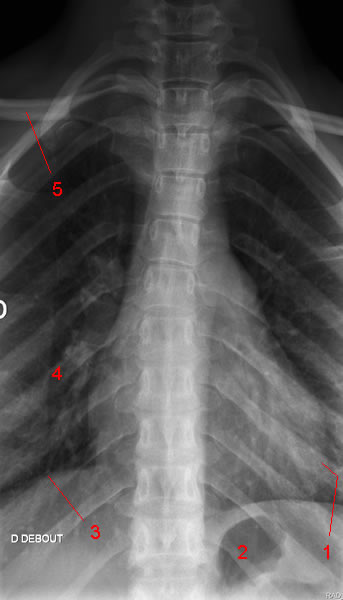
Image 1. Thoracic Spine X-ray: AP projection. 1, Left ventricle. 2, Gastric bubble. 3, Right hemidiaphragm. 4, Posterior rib (right side). 5, Clavicle.
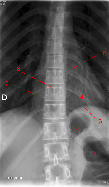
Image 2. Thoracic Spine X-ray: AP projection. 1, Gas in colon (Splenic flexure). 2, Gastric bubble. 3, Left hemidiaphragm. 4, Posterior rib. 5, Pedicle. 6, Spinous process. 7, Transverse process.
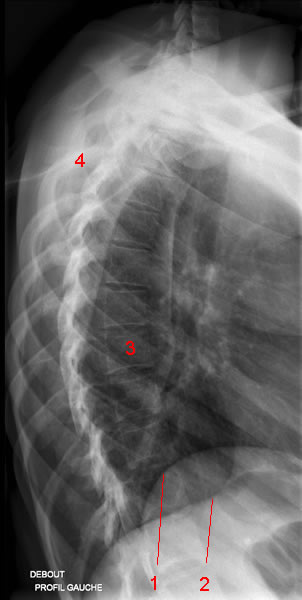
Image 3. Thoracic Spine X-ray: Lateral view. 1, Right hemidiaphragm. 2, Left hemidiaphragm. 3, Vertebral body. 4, Posterior rib.
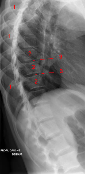
Image 4 of 4. Thoracic Spine X-ray: Lateral view. 1, Posterior rib. 2, Vertebral body. 3, Intervertebral disc space.
EspañolFrançais