-
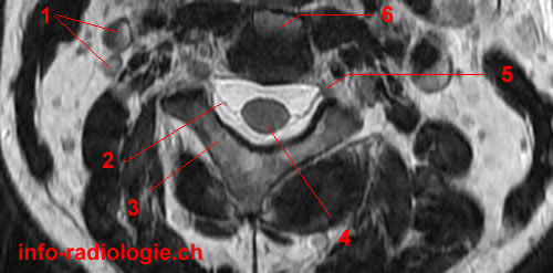
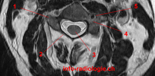
Image 1. MRI of the Cervical Spine, axial T2-weighted image. 1, Jugular vein and Carotid artery. 2, Nerve root. 3, Lamina. 4, Spinal cord. 5, Intervertebral foramen. 6, Inferior endplate, C2.
-
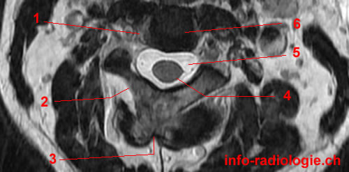
Image 2. MRI of the Cervical Spine, axial T2-weighted image. 1, Unciform process (C3). 2, Lamina (C2). 3, Spinous process (C2). 4, Spinal cord. 5, Cerebrospinal fluid. 6, Intervertebral disc space (C2-C3).
-
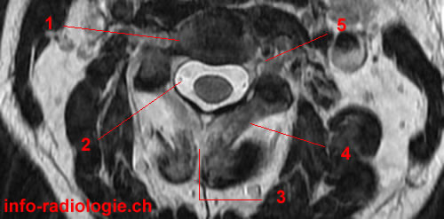
Image 3. MRI of the Cervical Spine, axial T2-weighted image. 1, Vertebral body, C3. 2, Cerebrospinal fluid. 3, Spinous process (C2). 4, Lamina. 5, Intervertebral foramen.
-
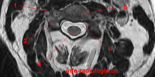
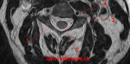
Image 4. MRI of the Cervical Spine, axial T2-weighted image. 1, Transverse foramen (C3). 2, Pedicle (C3). 3, Spinal cord. 4, Cerebrospinal fluid. 5, Vertebral body (C3).
-
Image 5. MRI of the Cervical Spine, axial T2-weighted image. 1, Transverse process (C3). 2, Lamina (C3). 3, Spinal cord. 4, Pedicle. 5, Transverse foramen (C3).
-
Image 6. MRI of the Cervical Spine, axial T2-weighted image. 1, Lamina (C3). 2, Spinal cord. 3, Transverse process (C3). 4, Vertebral artery.
-
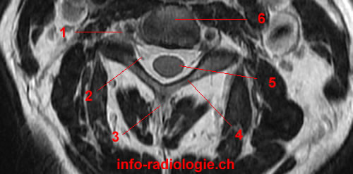
Image 7. MRI of the Cervical Spine, axial T2-weighted image. 1, Intervertebral foramen. 2, Posterior nerve root. 3, Spinous process (C3). 4, Lamina. 5, Spinal cord. 6, Vertebral body (C3).
-
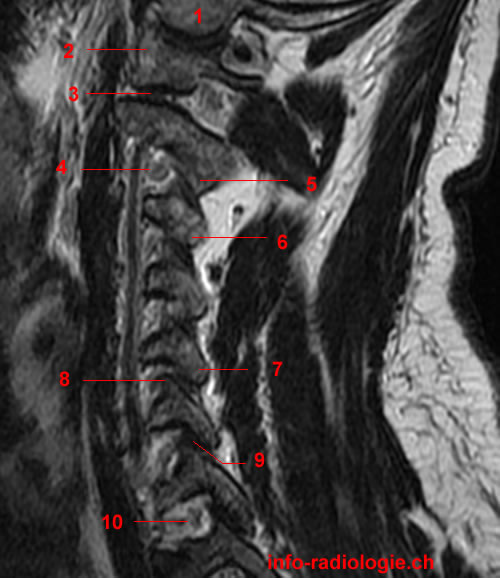
Image 8. MRI of the Cervical Spine, sagittal T2-weighted image. 1, Atlantooccipital joint. 2, Occipital condyle. 3, Vertebral artery. 4, Lateral mass of C1. 5, Inferior articular process of C2. 6, Superior articular process of C3. 7, Inferior articular process of C3. 8, Superior articular process of C6. 9, Inferior articular process of C6.
-
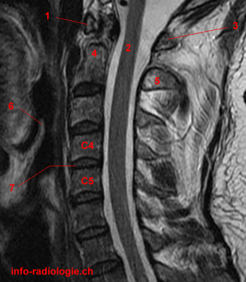
Image 9. MRI of the Cervical Spine, sagittal T2-weighted image. 1, Occipital condyle. 2, Lateral mass, C1. 3, Atlanto-axial joint. 4, Intervertebral foramen. 5, Inferior articular process, C2. 6, Inferior articular process C3. 7, Inferior articular process of C5. 8, Superior articular process of C6. 9, Facet joint. 10, Intervertebral foramen.
-
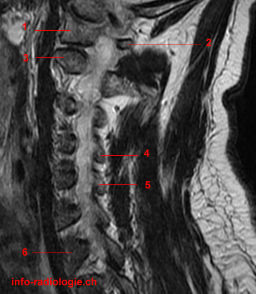
Image 10. MRI of the Cervical Spine, sagittal T2-weighted image. 1, Lateral mass, C1. 2, Posterior arch of C1. 3, Junction transverse process/vertebral body, C2 (axis). 4, Lamina (C4). 5, Lamina (C5). 6, Vertebral body, C7.
-
Image 11. MRI of the Cervical Spine, sagittal T2-weighted image. 1, Lateral mass of C1 (atlas). 2, Posterior arch of C1. 3, Vertebral foramen with cerebrospinal fluid. 4, Spinous process of l'axis.
-
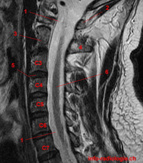
Image 12. MRI of the Cervical Spine, sagittal T2-weighted image. 1, Vertebral foramen - Cerebrospinal fluid. 2, Posterior arch of C1. 3, Vertebral body, C2. 4, Spinous process of C2. 5, Intervertebral disc space. 6, Spinal cord.
-
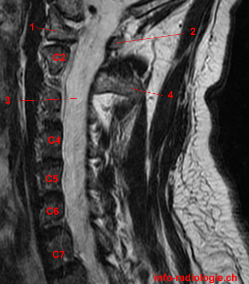
Image 13. MRI of the Cervical Spine, sagittal T2-weighted image. 1, Anterior arch of C1. 2, Spinal cord. 3, Posterior arch of l'Atlas (C1). 4, Ondotoid process, (C2). 5, Spinous process of C2. 6, Epiglottic cartilage. 7, Intervertebral disc space.
-
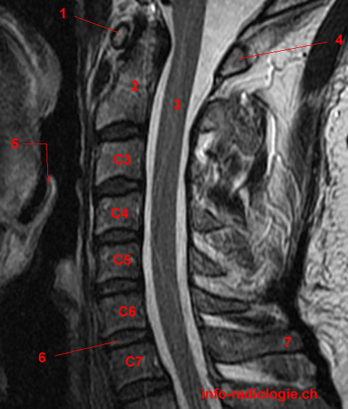
Image 14 of 14. MRI of the Cervical Spine, sagittal T2-weighted image. 1, Anterior arch of l'Atlas (C1). 2, Ondotoid process de l'axis (C2). 3, Spinal cord. 4, Posterior arch of C1. 5, Epiglottic cartilage. 6, Intervertebral disc space. 7, Spinous process of C7.