-
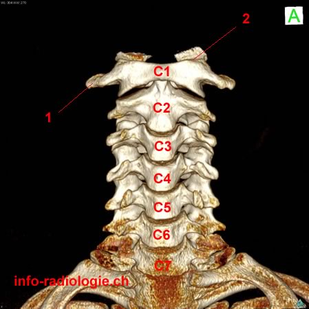
Image 1. Cervical spine: anterior view. 1, Transverse process. 2, Atlantooccipital joint. C1, Vertebral body of C1. .... C7, Vertebral body of C7.
-
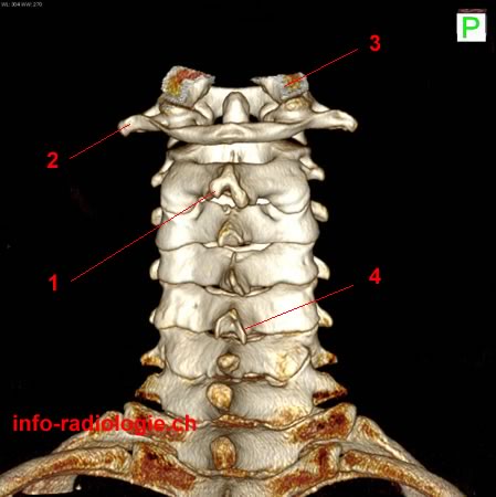
Image 2. Cervical spine: posterior view. 1, Spinous process of C2 (axis). 2, Transverse process of C1 (atlas). 3, Occipital condyle. 4, Spinous process of C5.
-
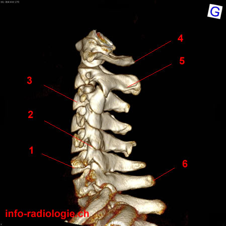
Image 3. Cervical spine: Lateral view. 1, Vertebral body of C6. 2, Zygapophysial joint. 3, Vertebral body of C3. 4, Posterior tubercle of C1. 5, Spinous process of C2. 6, Spinous process of C7.
-
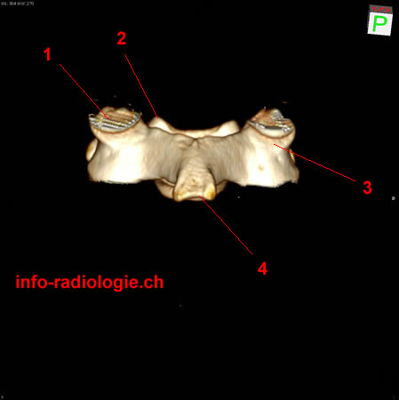
Image 4. Superior cervical spine (magnification): anterior view, without C1. 1, Transverse process of C2. 2, Odontoid process. 3, Superior articular facet. C3, Vertebral body of C3. C4, Vertebral body of C4.
-
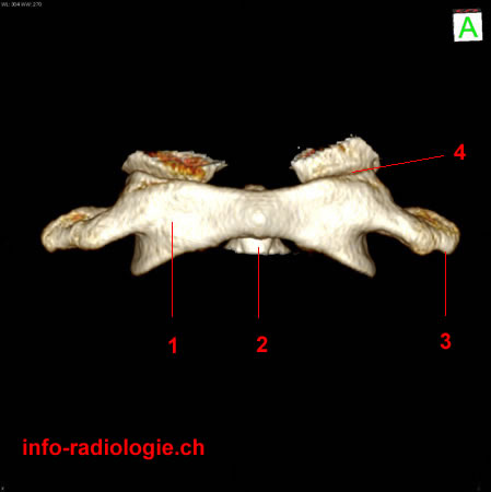
Image 5. C1 vertebra or Atlas: anterior view. 1, Lateral mass of C1. 2, Odontoid process. 3, Transverse process. 4, Atlantooccipital joint.
-
Image 6. C1 vertebra or Atlas: posterior view. 1, Apex of odontoid process. 2, Atlantooccipital joint. 3, Transverse process. 4, Posterior tubercle. 5, Posterior arch.
-
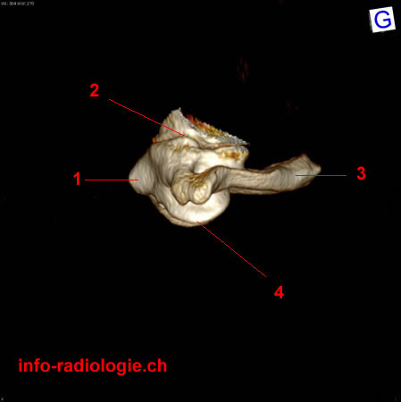
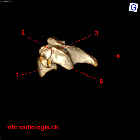
Image 7. C1 vertebra or atlas: lateral view. 1, Anterior tubercle. 2, Atlantooccipital joint. 3, Posterior arch of C1. 4, Atlanto-axial joint.
-
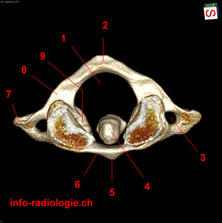
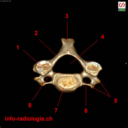
Image 8. C1 vertebra or atlas: superior view. 1, Vertebral foramen. 2, Posterior arch. 3, Transverse foramen. 4, Odontoid process. 5, Anterior tubercle. 6, Anterior arch. 7, Transverse process. 8, Canal of vertebral artery. 9, Lateral masse.
-
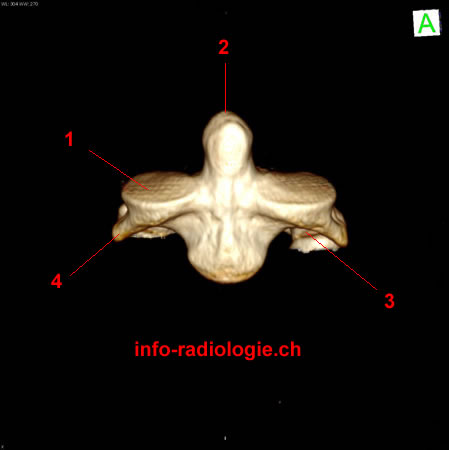
Image 9. C2 vertebra or Axis: anterior view. 1, Articular facet. 2, Dens of axis. 3, Inferior articular process. 4, Transverse process.
-
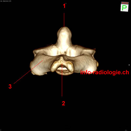
Image 10. C2 vertebra or Axis: posterior view. 1, Dens of axis. 2, Spinous process. 3, Posterior arch.
-
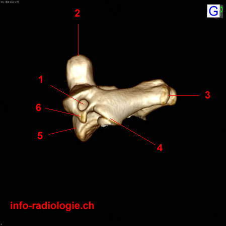
Image 11. C2 vertebra or Axis: lateral view. 1, Transverse foramen. 2, Dens (apex). 3, Spinous process. 4, Inferior articular process. 5, Vertebral body. 6, Transverse process .
-
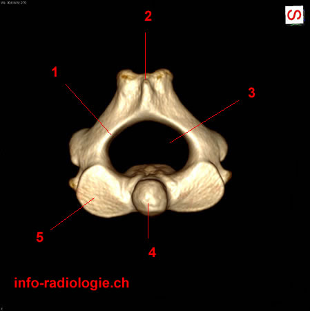
Image 12. C2 vertebra or Axis: superior view. 1, Posterior arch. 2, Spinous process. 3, Vertebral foramen. 4, Odontoid process. 5, Articular facet.
-
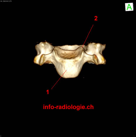
Image 13. Cervical vertebra (but C1 and C2): anterior view. 1, Vertebral body. 2, Unciform process.
-
Image 14. Cervical vertebra (but C1 and C2): posterior view. 1, Articular facet. 2, Unciform process. 3, Superior articular process. 4, Spinous process.
-
Image 15. Cervical vertebra (but C1 and C2): lateral view. 1, Vertebral body. 2, Superior articular process. 3, Articular facet. 4, Spinous process. 5, Inferior articular process.
-
Image 16 of 16. Cervical vertebra (but C1 and C2): superior view. 1, Superior articular facet. 2, Lamina. 3, Spinous process. 4, Vertebral foramen. 5, Transverse process. 6, Transverse foramen. 7, Vertebral body. 8, Pedicle.