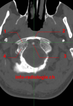
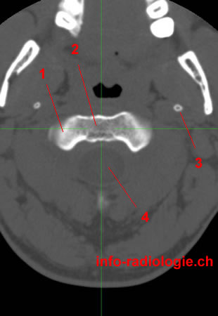
CT of the Craniocervical junction, axial reconstruction. Level 1. Image 1.
1, Atlantooccipital joint. 2, Anterior arch (Atlas-C1). 3, Dens (apex). 4, Occipital condyle.

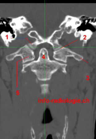
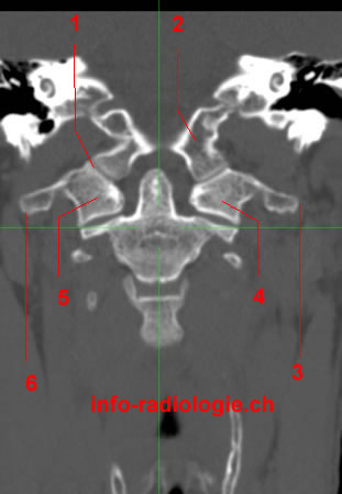
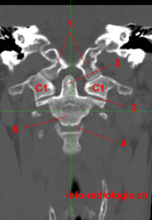
CT of the Craniocervical junction, coronal reconstruction. Level 1. Image 2
1, Occipital condyle. 2, Atlantooccipital joint. 3, Transverse process (Atlas-C1). 4, Dens (Axis-C2). 5, Lateral mass (Atlas-C1).

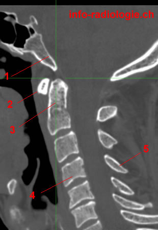
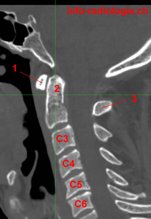
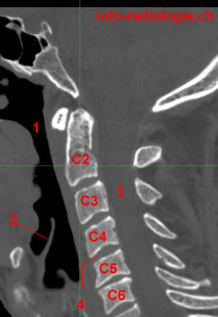
CT of the Craniocervical junction, sagittal reconstruction. Level 1. Image 3. 1, Clivus. 2, Anterior arch (Atlas-C1). 3, Dens (Axis-C2). 4, Vertebral body C4. 5, Spinous process C4.

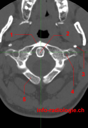
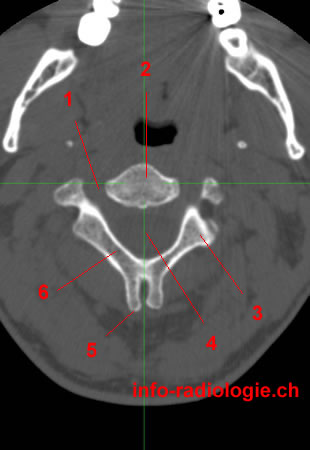
CT of the Craniocervical junction, axial reconstruction. Level 2. Image 1. 1, Anterior arch (Atlas-C1). 2, Dens (Axis-C2). 3, Styloid process. 4, Transverse foramen. 5, Posterior arch (Atlas-C1).

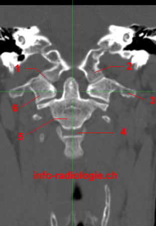
CT of the Craniocervical junction, coronal reconstruction. Level 2. Image 2. 1, Atlantooccipital joint. 2, Occipital condyle. 3, Transverse process (C1). 4, Intervertebral space (disc) C2-C3. 5, Vertebral body (Axis-C2). 6, Lateral mass (Atlas-C1).

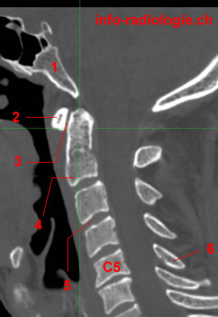
CT of the Craniocervical junction, sagittal reconstruction. Level 2. Image 3 1, Clivus. 2, Anterior arch (Atlas-C1). 3, Atlantoaxial joint. 4, Axis (C2). 5, Intervertebral space (disc) C3-C4. 6, Spinous process de C5.

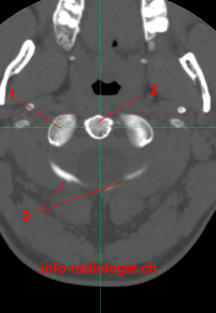
CT of the Craniocervical junction, axial reconstruction. Level 3. Image 1. 1, Inferior articular surface (Atlas-C1). 2, Dens (Axis-C2). 3, Posterior arch (Atlas-C1).

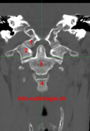
CT of the Craniocervical junction, coronal reconstruction. Level 3. Image 2. 1, Occipital condyle. 2, Atlas (C1). 3, Axis (C2). 4, Vertebral body (C3).

CT of the Craniocervical junction, sagittal reconstruction. Level 3. Image 3. 1, Anterior arch (Atlas-C1) 2, Dens (Axis-C2). 3, Spinous process (C2).

CT of the Craniocervical junction, axial reconstruction. Level 4. Image 1. 1, Superior articular surface (Axis-C2). 2, Body (Axis-C2). 3, Styloid process. 4, Vertebral foramen.

CT of the Craniocervical junction, coronal reconstruction. Level 4. Image 2 1, Atlantooccipital joint. 2, Occipital condyle. 3, Transverse process (Axis-C1) - left side. 4, Lateral mass of C1 (left side). 5, Lateral mass of C1 (right side).6, Transverse process (Axis-C1) - right side.

CT of the Craniocervical junction, sagittal reconstruction. reconstruction sagittale. Level 4. Image 3 1, Clivus. 2, Anterior arch (Atlas-C1). 3, Spinous process de C3.

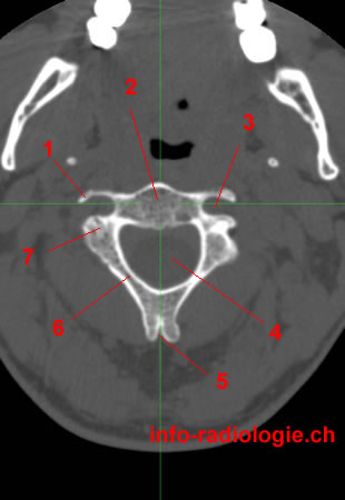
CT of the Craniocervical junction, axial reconstruction. Level 5. Image 1 1, Transverse process (C2). 2, Vertebral body (Axis-C2). 3, Transverse foramen (C2). 4, Vertebral foramen. 5, Spinous process (C2). 6, Lamina. 7, Pedicle.

CT of the Craniocervical junction, coronal reconstruction. Level 5. Image 2. 1, Occipital condyle. 2, Dens (Axis-C1) 3, Atlantoaxial joint. 4,Intervertebral space (disc) C2-C3. 5, Corps (Axis-C2).

CT of the Craniocervical junction, sagittal reconstruction. Level 5. Image 3. 1, Clivus. 2, Vertebral foramen. 3, Spinous process of C5.

CT of the Craniocervical junction, axial reconstruction. Level 6. Image 1. 1, Intervertebral foramen. 2, Vertebral body (Axis-C2). 3, Inferior articular process (C2). 4, Vertebral foramen. 5, Bifid spinous process (Axis-C2). 6, Lamina.

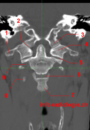
CT of the Craniocervical junction, coronal reconstruction. Level 6. Image 2. 1, Transverse process of C1. 2, Articulation atlanto-occipital. 3, Occipital condyle. 4, Lateral mass of C1. 5, Atlantoaxial joint. 6,Intervertebral space (disc) C2-C3. 7, Vertebral body C3. 8, Tip of transverse process de C3. 9, Transverse foramen (C2).

. CT of the Craniocervical junction, sagittal reconstruction. Level 6 de 6. Image 3 (Last level-last image) 1, Pharynx. 2, Epiglotic cartilage. 3, Vertebral foramen. 4,Intervertebral space (disc) C4-C5.