Sacrum and coccyx

This photo gallery presents the anatomy of sacrum and coccyx by means of 3D-reconstructions, axial, sagittal and coronal reconstructions obtained from a scan of pelvis.

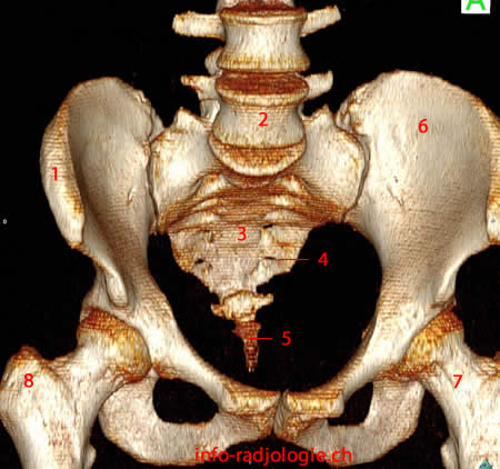
Pelvis, anterior view, 3D reconstruction (CT-scan). Image 1.1, Iliac crest. 2, Lumbar vertebre. 3, Sacrum. 4, Ventral sacral foramen. 5, Coccyx. 6, Iliac wing. 7, Femoral neck (Left side). 8, Greater trochanter (fémur/right side).

Sacrum and coccyx (CT-scan). Image 1
  • Pelvis, anterior view, 3D reconstruction (CT-scan). Image 1.  1, Iliac crest. 2, Lumbar vertebre. 3, Sacrum. 4, Ventral sacral foramen. 5, Coccyx. 6, Iliac wing. 7, Femoral neck (Left side). 8, Greater trochanter (fémur/right side).

  • Pelvis, posterior view, 3D reconstruction (CT-scan). Image 2.   1, Iliac wing. 2, Median crest of sacrum. 3, Lateral sacral crest. 4, Intermediate sacral crest. 5, Dorsal sacral foramen.

  • Sacrum, CT-scan, coronal oblique reconstruction. Image 3.   1, Geode (Lumbar vertebre). 2, Sacral ala. 3, Sacroiliac joint (Left side). 4, Ventral sacral foramen.

  • Sacrum, CT-scan, coronal oblique reconstruction. Image 4.   1, Lumbar vertebre. 2, Sacral canal. 3, Ventral sacral foramen.

  • Sacrum, CT-scan, axial reconstruction. Image 5  1, Iliac crest. 2, Sacroiliac joint (right side). 3, Dorsal sacral foramen. 4, Ventral sacral foramen.

  • Sacrum, CT-scan, axial reconstruction. Image 6.  1, Iliac crest. 2, Ventral sacral foramen. 3, Sacral canal. 4, Sacroiliac joint (Left side).

  • Sacrum, CT-scan, axial reconstruction. Image 7.  1, Ventral sacral foramen. 2, Dorsal sacral foramen.

  • Sacrum, CT-scan, sagittal reconstruction. Image 8.  1, Vertebral body (lumbar spine). 2, Intervertebral disc. 3, Sacral canal. 4, Median crest.

  • Sacrum, CT-scan, sagittal reconstruction. Image 9  1, Vertebral body S1. 2, Ventral sacral foramen. 3, Ventral sacral foramen. 4, Dorsal sacral foramen.

  • Sacrum, CT-scan, 3D reconstruction, anterior view. Image 10.  1, Ventral sacral foramen. 2, Lumbar vertebre. 3, Promontory. 4, Sacroiliac joint (Left side).

  • Sacrum, CT-scan, 3D reconstruction, superior and anterior view. Image 11.  1, Sacral ala. 2, Promontory. 3, Sacrococcygeal joint. 4, Transverse ridge.

  • Sacrum, reconstruction 3D (scanner), anterior and oblique view. Image 12 of 12.  1, Sacral ala. 2, Superior articular process, S1. 3, Sacroiliac joint. 4, Sacrococcygeal joint. 5, Coccyx. 6, Promontory.