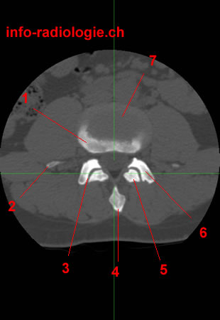
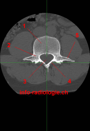
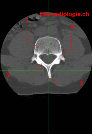
CT of lumbar Spine, axial reconstruction. Level 1. Image 1. 1, Vertebral body (L4). 2, Transverse process. 3, Facet joint. 4, Spinous process. 5, Inferior articular process L3. 6, Superior articular process of L4. 7, Intervertebral disc L3-L4.

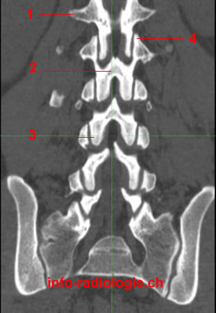
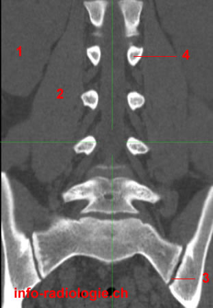
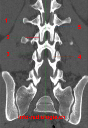
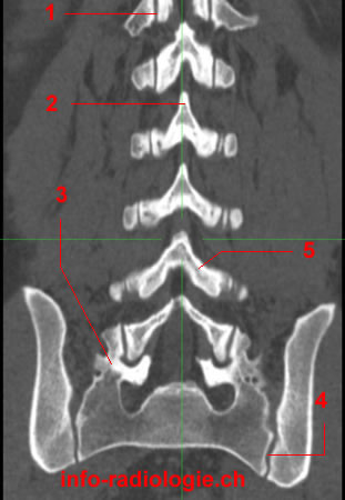
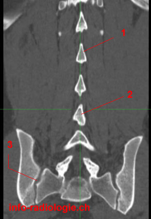
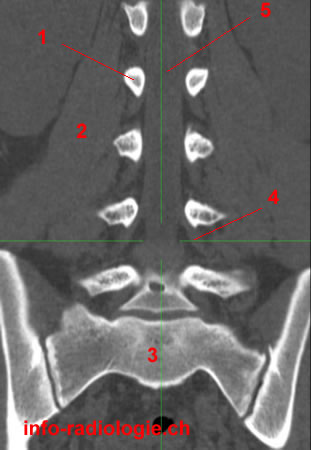
CT of lumbar Spine, coronal reconstruction. Level 1. Image 2. 1, Transverse process de L1. 2, Lamina. 3, Inferior articular process of L3. 4, Superior articular process of L2.

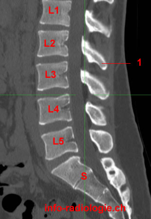
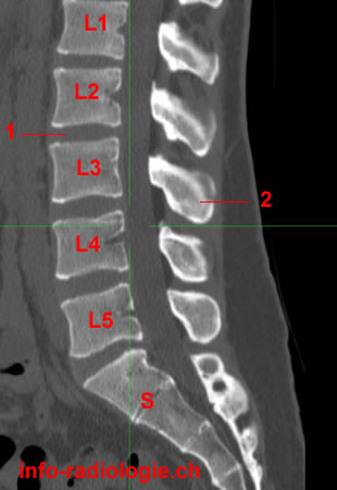
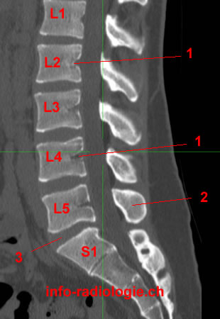
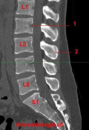
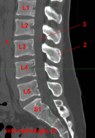
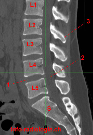
CT of lumbar Spine, sagittal reconstruction.Level 1. Image 3. 1, Spinous process de L2. S, Sacrum. L1, Vertebral body, L1. L2, Vertebral body, L2. etc.

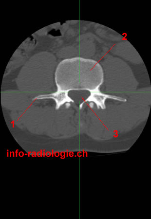
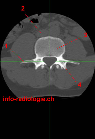
CT of lumbar Spine, axial reconstruction. Level 2. Image 1. 1, Transverse process. 2, Vertebral body (L4). 3, Vertebral foramen.

CT of lumbar Spine, coronal reconstruction. Level 2. Image 2. 1, Right kidney. 2, Psoas muscle (right side). 3, Sacroiliac joint. 4, Pedicle (left side - L2).

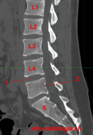
CT of lumbar Spine, sagittal reconstruction. Level 2. Image 3. 1, Intervertebral disc space. 2, Spinous process (L3). S, Sacrum. L1, Vertebral body, L1. L2, Vertebral body, L2. etc.

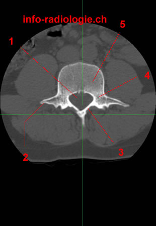
CT of lumbar Spine, axial reconstruction. Level 3. Image 1.1, Transverse process. 2, Inferior vena cava. 3, Vertebral body (L4). 4, Superior articular process.

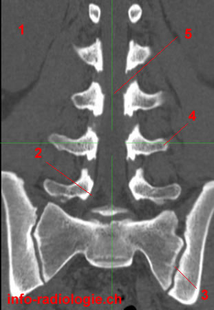
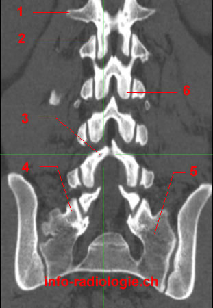
CT of lumbar Spine, coronal reconstruction. Level 3. Image 2. 1, Right kidney. 2, Nerve root. 3, Sacroiliac joint (left side). 4, Transverse process of L4. 5, Vertebral foramen.

CT of lumbar Spine, sagittal reconstruction. Level 3. Image 3. 1, Intervertebral disc space. 2, Spinous process of L3. S, Sacrum. L1, Vertebral body, L1. L2, Vertebral body, L2. etc.

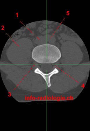
CT of lumbar Spine, axial reconstruction. Level 4. Image 1. 1, Vertebral body (L4). 2, Pedicle. 3, Vertebral foramen. 4, Lamina. 5, Transverse process.

CT of lumbar Spine, coronal reconstruction. Level 4. Image 2. 1, Transverse process of L2. 2, Facet joint. 3, Superior articular process of L4. 4, Inferior articular process L3. 5, Lamina.

CT of lumbar Spine, sagittal reconstruction. Level 4. Image 3. 1, Basivertebral vein. 2, Spinous process of L5. 3, Intervertebral disc space L5-S1. S, Sacrum. L1, Vertebral body, L1. L2, Vertebral body, L2. etc.

CT of lumbar Spine, axial reconstruction. Level 5. Image 1. 1, Vertebral foramen. 2, Transverse process. 3, Lamina. 4, Pedicle. 5, Vertebral body (L4).

CT of lumbar Spine, coronal reconstruction. Level 5. Image 2. 1, Facet joint. 2, Spinous process of L2. 3, Superior articular process (sacrum). 4, Sacroiliac joint. 5, Lamina.

CT of lumbar Spine, sagittal reconstruction. Level 5. Image 3. 1, Basivertebral vein. 2, Spinous process of L3. S, Vertebral body, S1. L1, Vertebral body, L1. L2, Vertebral body, L2. etc.

CT of lumbar Spine, axial reconstruction. Level 6. Image 1 1, Inferior vena cava. 2, Psoas muscle (right side). 3, Spinous process. 4, Lamina. 5, Vertebral body (L4). 6, Aorta.

CT of lumbar Spine, coronal reconstruction. Level 6. Image 2. 1, Transverse process of L1. 2, Superior articular process of L2. 3, Lamina. 4, Superior articular process (S1). 5, Sacral ala. 6, Inferior articular process L2.

CT of lumbar Spine, sagittal reconstruction. Level 6. Image 3. 1, Intervertebral disc space. 2, Basivertebral vein. S, Sacrum. L1, Vertebral body, L1. L2, Vertebral body, L2. etc.

CT of lumbar Spine, axial reconstruction. Level 7. Image 1. 1, Vertebral body (L4). 2, Lamina. 3, Spinous process. 4, Pedicle.

CT of lumbar Spine, coronal reconstruction. Level 7. Image 2. 1, Spinous process of L2. 2, Spinous process of L4. 3, Sacroiliac joint.

CT of lumbar Spine, sagittal reconstruction. Level 7. Image 3. 1, Aorta. 2, Spinous process de L3. 3, Vertebral foramen. S, Sacrum. L1, Vertebral body, L1. L2, Vertebral body, L2. etc.

CT of lumbar Spine, axial reconstruction. Level 8 (last level). Image 1. 1, Inferior vena cava. 2, Psoas muscle. 3, Nerve root. 4, Intervertebral foramen. 5, Aorta.

CT of lumbar Spine, coronal reconstruction. Level 8 (last level). Image 2. 1, Pedicle (L2). 2, Psoas muscle. 3, Os sacrum. 4, Nerve root. 5, Vertebral foramen.

CT of lumbar Spine, sagittal reconstruction. Level 8 (last level). Image 3. 1, Intervertebral disc space. 2, Vertebral foramen. 3, Spinous process de L2. S, Sacrum. L1, Vertebral body, L1. L2, Vertebral body, L2. etc.