CT scan of the middle ear (anatomy)

This page presents the anatomy of middle ear by means of CT-scan.

You have three different galleries of images depicting the anatomy of the middle ear:
• level 1 to 6 (axial, coronal, sagittal reconstruction for the same level . A set of 3 images correspond to a given level and the green cross indicates the reference planes for each image of this level .)
• Serie of successive coronal reconstructions
• Serie of successive sagittal reconstructions

level 1 to 6


  • CT of middle ear- Image 1
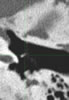
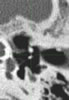
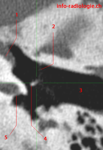
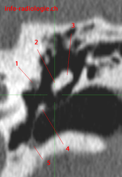
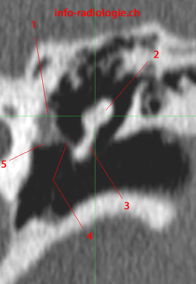
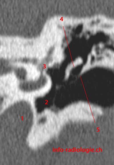
    CT of middle ear and ossicles, level 1, axial reconstruction (magnification). 1, Tensor tympani muscle. 2, Tympanic membrane. 3, External auditory canal. 4, Manubrium of malleus. 5, Round window.
  • CT of middle ear- Image 2
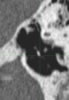
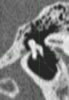
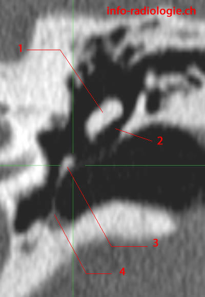
    CT of middle ear and ossicles, level 1, coronal reconstruction (magnification). 1, Body of incus. 2, Prussak Space. 3, Manubrium of malleus. 4, Tympanic annulus.
  • CT of middle ear- Image 3
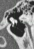
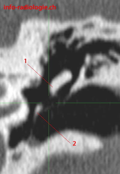
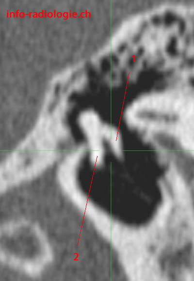
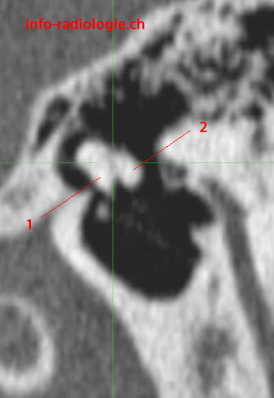
    CT of middle ear and ossicles, level 1, sagittal reconstruction (magnification). 1, Malleus.
  • CT of middle ear- Image 4
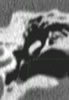
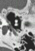
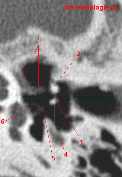
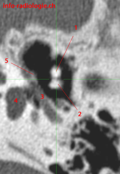
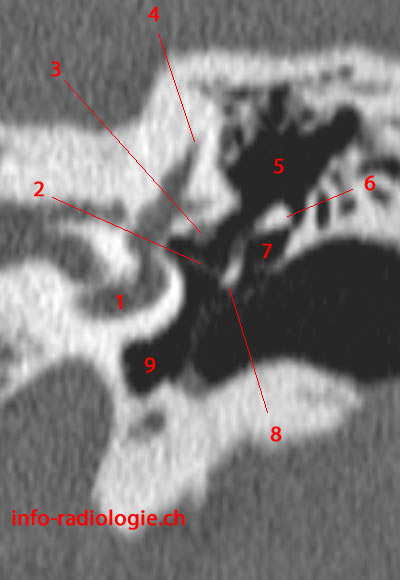
    CT of middle ear and ossicles, level 2, axial reconstruction (magnification). 1, Tensor tympani muscle. 2, Malleus. 3, External auditory canal. 4, Incus. 5, Recess of the facial nerve. 6, Sinus tympani. 7, Pyramidal eminence.
  • CT of middle ear- Image 5
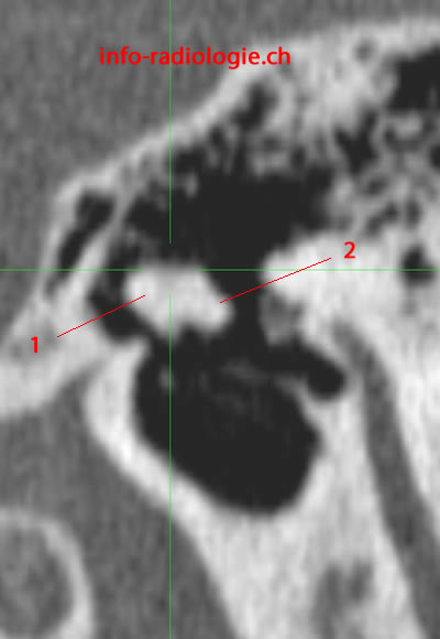
    CT of middle ear and ossicles, level 2, coronal reconstruction (magnification). 1, Incus. 2, Malleus.
  • CT of middle ear- Image 6
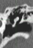
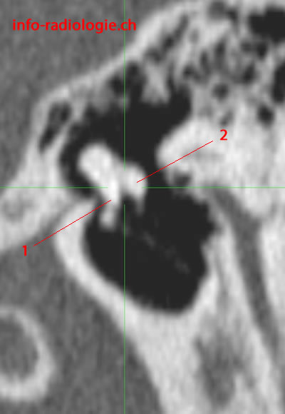
    CT of middle ear and ossicles, level 2, sagittal reconstruction (magnification). 1, Malleus. 2, Incus.
  • CT of middle ear- Image 7
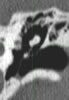
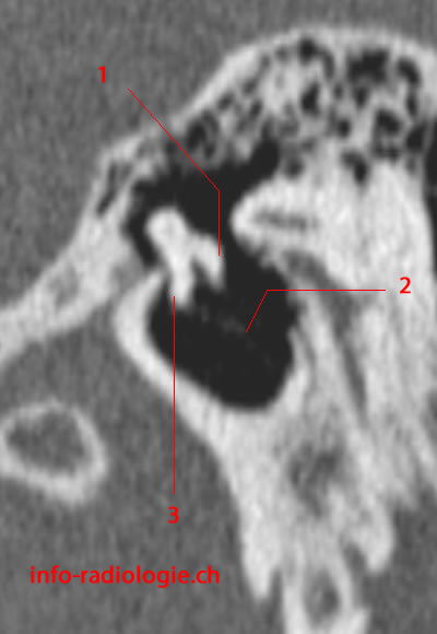
    CT of middle ear and ossicles, level 3, axial reconstruction (magnification). 1, Tensor tympani Tendon & muscle. 2, Malleus. 3, Incus. 4, Stapedius muscle. 5, Stapes. 6, Base of stapes.
  • CT of middle ear- Image 8
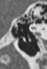
    CT of middle ear and ossicles, level 3, coronal reconstruction (magnification). 1, Facial nerve canal. 2, Malleus. 3, Incus. 4, Malleus (manubrium). 5, Tympanic annulus.
  • CT of middle ear- Image 9
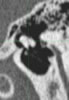
    CT of middle ear and ossicles, level 3, sagittal reconstruction (magnification). 1, Incus. 2, Malleus.
  • CT of middle ear- Image 10
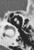
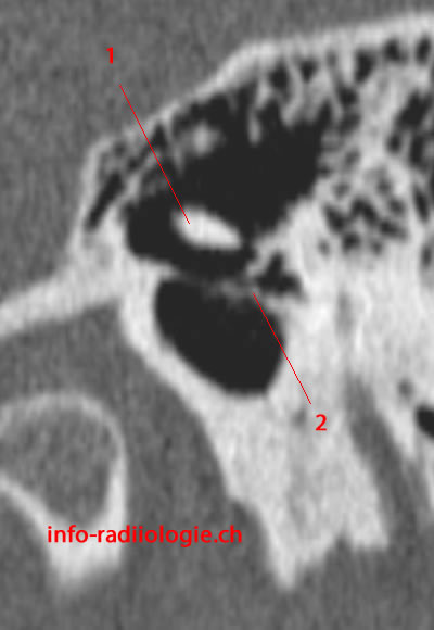
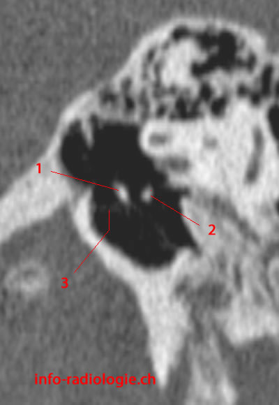
    CT of middle ear and ossicles, level 4, axial reconstruction (magnification). 1, Malleus. 2, Incus. 3, Facial nerve canal. 4, Vestibule. 5, Cochlear process.
  • CT of middle ear- Image 11
    CT of middle ear and ossicles, level 4, coronal reconstruction (magnification). 1, Facial nerve canal. 2, Malleus. 3, Incus. 4, Malleus (manubrium). 5, Tympanic annulus.
  • CT of middle ear- Image 12
    CT of middle ear and ossicles, level 4, sagittal reconstruction (magnification). 1, Malleus. 2, Incus.
  • CT of middle ear- Image 13
    CT of middle ear and ossicles, level 5, axial reconstruction (magnification). 1, Head of malleus. 2, Prussak Space. 3, Koerner septum. 4, Mastoid (antrum). 5, Incus (body).
  • CT of middle ear- Image 14
    CT of middle ear and ossicles, level 5, coronal reconstruction (magnification). 1, Facial nerve canal. 2, Incus. 3, Malleus. 4, Tensor tympani Tendon & muscle. 5, Cochlear process.
  • CT of middle ear- Image 15
    CT of middle ear and ossicles, level 5, sagittal reconstruction (magnification). 1, Malleus. 2, Incus.
  • CT of middle ear- Image 16
    CT of middle ear and ossicles, level 6 of 6, axial reconstruction (magnification). 1, Anterior epitympanic recess. 2, Head of malleus. 3, Incudo-mallear joint. 4, Prussak Space. 5, Incus (body). 6, Koerner septum. 7, Mastoid (antrum). 8, Lateral semicircular canal. 9, Epitympanum.
  • CT of middle ear- Image 17
    CT of middle ear and ossicles, level 6 of 6, coronal reconstruction (magnification). 1, Facial nerve canal. 2, Incus. 3, Malleus. 4, Tensor tympani Tendon & muscle. 5, Cochlear process.
  • CT of middle ear- Image 18
    CT of middle ear and ossicles,level 6 of 6 (last image), sagittal reconstruction (magnification). 1, Malleus. 2, Incus.
Oreille moyenne,level 1, axial reconstruction (magnification). 1, Tensor tympani muscle. 2, Tympanic membrane. 3, External auditory canal. 4, Manubrium of malleus. 5, Round window.
CT of middle ear- Image 1

Serie of successive coronal reconstructions


Image 1. CT of middle ear, coronal reconstruction (magnification). 1, Internal carotid artery. 2, Cochlea (1st turn). 3, Cochlea (2nd turn). 4, Malleus (head). 5, Tympanic membrane.
Coupe coronale de l'oreille moyenne. Image 1
  • Image 1. CT of middle ear, coronal reconstruction (magnification). 1, Internal carotid artery. 2, Cochlea (1st turn). 3, Cochlea (2nd turn). 4, Malleus (head). 5, Tympanic membrane.
  • Image 2. CT of middle ear, coronal reconstruction (magnification). 1, Cochlea. 2, Malleus (head). 3, Incus. 4, Scutum. 5, Malleus (manubrium). 6, Tympanic membrane. 7, Internal carotid artery.
  • Image 3. CT of middle ear, coronal reconstruction (magnification). 1, Internal carotid artery. 2, Cochlea (1st turn). 3, Cochlea (2nd turn). 4, Labyrinthine segment of the facial nerve canal. 5, Malleus (head). 6, Incus. 7, Malleus (manubrium). 8, Tensor tympani Tendon & muscle. 9, Cochlear process.
  • Image 4. CT of middle ear, coronal reconstruction (magnification). 1, Internal carotid artery. 2, Hypothympanum. 3, Vestibule. 4, Incus. 5, Scutum.
  • Image 5 of 5. CT of middle ear, coronal reconstruction (magnification). 1, Cochlea. 2, Stapes. 3, Facial nerve (pars tympanic). 4, Superior semicircular canal. 5, Epitympanum. 6, Incus/processus court. 7, Prussak Space.8, Incus/long process. 9, Hypotympan.

Series of successive sagittal reconstructions


Image 1. CT of middle ear, sagittal reconstruction (magnification). 1, Malleus. 2, Scutum.
Coupe sagittale de l'oreille moyenne. Image 1
  • Image 1. CT of middle ear, sagittal reconstruction (magnification). 1, Malleus. 2, Scutum.
  • Image 2. CT of middle ear, sagittal reconstruction (magnification). 1, Incus. 2, Tympanic membrane. 3, Malleus.
  • Image 3. CT of middle ear, sagittal reconstruction (magnification). 1, Malleus. 2, Incus. 3, Tympanic membrane.
  • Image 4 of 4. CT of middle ear, sagittal reconstruction (magnification). 1, Malleus. 2, Incus. 3, Tympanic membrane.

Reference

• Harnsberger HR, Osborn AG, Ross JS, Moore KR, Salzman KL, Carrasco CR, Halmiton BE, Davidson HC, Wiggins RH. Diagnostic and Surgical Imaging Anatomy: Brain, Head and Neck, Spine. 3rd ed. Salt Lake City, Utah. Amirsys. 2007.
• Bourjat P, Veillon F. Imagerie radiologique tête et cou. Paris, Vigot. 1995.
• Gouazé A, Baumann JA, Dhem A. Sobota. Atlas d'Anatomie humaine. Tome 3. Système nerveux central, système nerveux autonome, organe des sens et peau, vaisseaux et nerfs périphériques. 1er éd. Paris, Maloine. 1977.
• Kahle W, Cabrol C. Anatomie. Tome 3: Système nerveux et organe des sens. 1er éd. Paris, Flammarion. 1979.