-
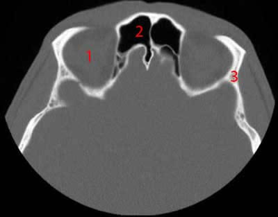
Image 1. CT Scan of the Paranasal Sinuses, axial reconstruction.
1, Orbital cavity. 2, Frontal sinus. 3, Sphenoid bone.
-
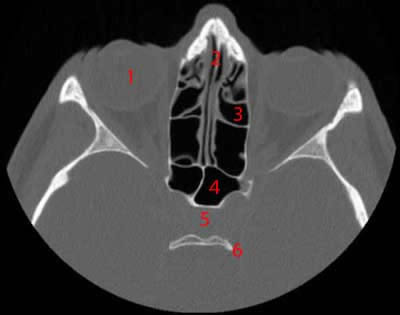
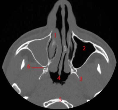
Image 2. CT Scan of the Paranasal Sinuses, axial reconstruction.
1, Globe. 2, Nasal septum. 3, Ethmoidal cells. 4, Sphenoidal sinus. 5, Pituitary gland. 6, Dorsum sellae.
-
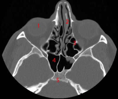
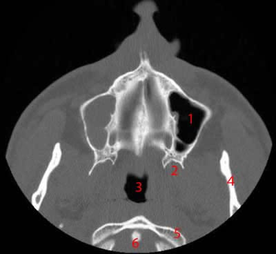
Image 3. CT Scan of the Paranasal Sinuses, axial reconstruction.
1, Globe. 2, Nasal septum. 3, Ethmoidal cells. 4, Sphenoidal sinus. 5, Clivus.
-
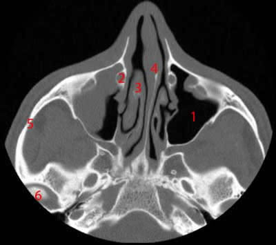
Image 4. CT Scan of the Paranasal Sinuses, axial reconstruction.
1, Maxillary sinus (left side). 2, Right nasolacrimal duct. 3, Turbinate. 4, Nasal septum. 5, Zygomatic arch. 6, Condylar process of mandible.
-
Image 5. CT Scan of the Paranasal Sinuses, axial reconstruction.
1, Nasal concha. 2, Maxillary sinus. 3, Lateral lamina of pterygoid process. 4, nasopharynx. 5, atlas. 6, Pterygomaxillary fissure.
-
Image 6. CT Scan of the Paranasal Sinuses, axial reconstruction.
1, Maxillary sinus. 2, Pterygoid process. 3, Nasopharynx. 4, Mandible. 5, Anterior arch of C1 (atlas). 6, Odontoid process (axis).
-
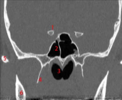
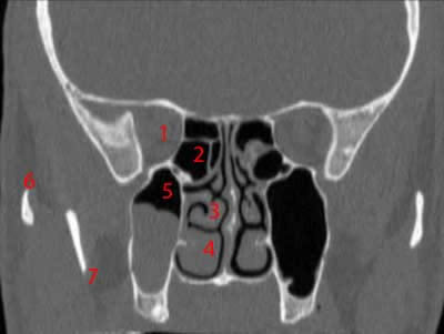
Image 7. CT Scan of the Paranasal Sinuses, Coronal reconstruction.
1, Anterior clinoid process. 2, Sphenoidal sinus. 3, Rhinopharynx. 4, Pterygoid hamulus. 5, Zygomatic arch. 6, Mandible.
-
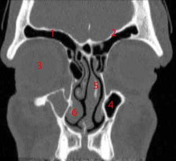
Image 8. CT Scan of the Paranasal Sinuses, Coronal reconstruction.
1, Sphenoidal sinus. 2, Middle nasal concha. 3, Inferior nasal concha. 4, Zygomatic arch. 5, Mandible.
-
Image 9. CT Scan of the Paranasal Sinuses, Coronal reconstruction.
1, Orbital cavity. 2, Ethmoidal cells. 3, Middle nasal concha. 4, Inferior nasal concha. 5, Maxillary sinus (right side). 6, Zygomatic arch. 7, Mandible.
-
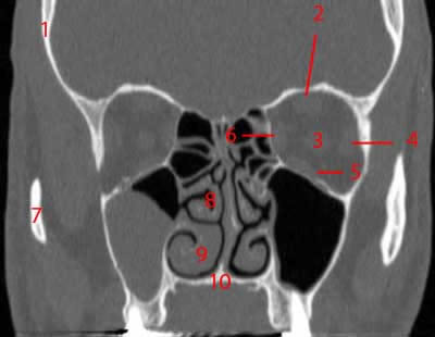
Image 10. CT Scan of the Paranasal Sinuses, Coronal reconstruction.
1, Frontal bone. 2, Superior rectus muscle. 3, Optic nerve. 4, Lateral rectus muscle. 5, Inferior rectus muscle. 6, Medial rectus muscle. 7, Zygomatic arch. 8, Middle nasal concha. 9, Inferior nasal concha. 10, Hard palate
-
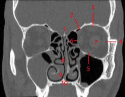
Image 11. CT Scan of the Paranasal Sinuses, Coronal reconstruction.
1, Crista galli. 2, Superior oblique muscle. 3, Superior rectus muscle. 4, Lateral rectus muscle 5, Inferior rectus muscle. 6, Medial rectus muscle. 7, Optic nerve. 8, Middle nasal concha. 9, Inferior nasal concha. 10, Hard palate.
-
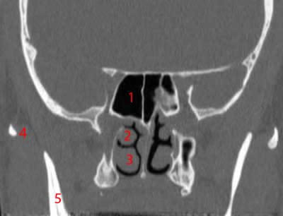
Image 12. CT Scan of the Paranasal Sinuses, Coronal reconstruction.
1, Frontal sinus. 2, Frontal bone. 3, Globe. 4, Maxillary sinus. 5, Nasal septum. 6, Inferior nasal concha.
-
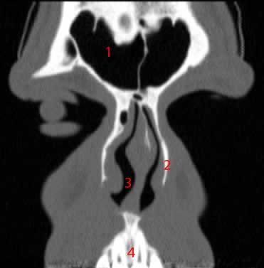
Image 13. CT Scan of the Paranasal Sinuses, Coronal reconstruction.
1, Frontal sinus. 2, Nasal bone. 3, Nasal cavity. 4, Alveolar arch.
-
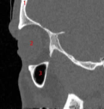
Image 14. CT Scan of the Paranasal Sinuses, sagittal reconstruction.
1, Frontal bone. 2, Orbital cavity. 3, Maxillary sinus.
-
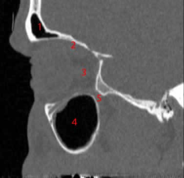
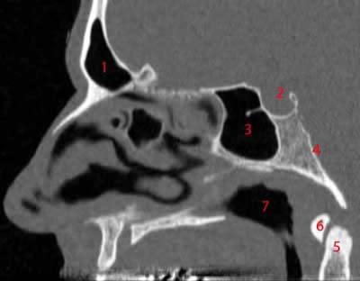
Image 15. CT Scan of the Paranasal Sinuses, sagittal reconstruction.
1, Frontal sinus. 2, Orbital cavity. 3, Optic nerve. 4, Maxillary sinus. 5, Inferior orbital fissure.
-
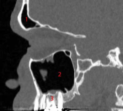
Image 16. CT Scan of the Paranasal Sinuses, sagittal reconstruction.
1, Frontal sinus. 2, Maxillary sinus. 3, Alveolar arch (maxilla).
-
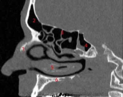
Image 17. CT Scan of the Paranasal Sinuses, sagittal reconstruction.
1, Nasal bone. 2, Frontal sinus. 3, Ethmoidal cells. 4, Sphenoidal sinus. 5, Inferior nasal concha. 6, Hard palate.
-
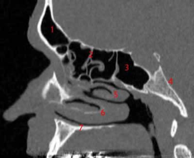
Image 18. CT Scan of the Paranasal Sinuses, sagittal reconstruction.
1, Frontal sinus. 2, Hypophyseal fossa. 3, Sphenoidal sinus. 4, clivus. 5, Odontoid process (C2 - axis). 6, Anterior arch of C1 (atlas). 7, rhinopharynx
-
Image 19 of 19. CT Scan of the Paranasal Sinuses, sagittal reconstruction.
1, Frontal sinus. 2, Ethmoidal cells. 3, Sphenoidal sinus. 4, Clivus. 5, Middle nasal concha. 6, Inferior nasal concha. 7, Hard palate.