This webpage presents the anatomical structures found on skull base CT.
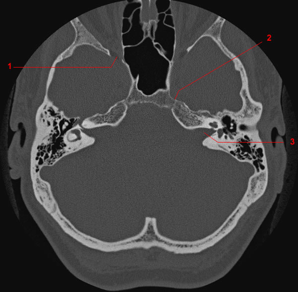
Image 1. CT anatomy of skull base. 1, Orbital cavity. 2, Superior orbital fissure. 3, Anterior clinoid process. 4, Mastoid air cells. 5, Internal occipital protuberance .
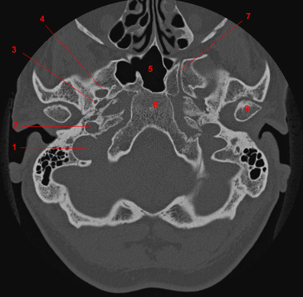
Image 2. CT anatomy of skull base. 1, Orbital cavity. 2, Superior orbital fissure. 3, Sphenoid sinus. 4, Optic canal. 5, Dorsum sellae. 6, Internal occipital protuberance .
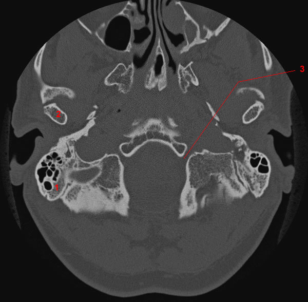
Image 3. CT anatomy of skull base. 1, Superior orbital fissure. 2, Petrooccipital fissure. 3, Internal auditory canal.
Image 4. CT anatomy of skull base. 1, Internal auditory canal. 2, Sphenoid sinus. 3, External auditory canal.
Image 5. CT anatomy of skull base. 1, Sigmoid sinus. 2, Jugular bulb. 3, Foramen rotondum. 4, Carotid canal (pars horizontal).
Image 6. CT anatomy of skull base. 1, Jugular bulb. 2, Carotid canal (pars horizontal). 3, Sphenoid sinus. 4, Foramen ovale. 5, Foramen spinosum. 6, Jugular foramen.
Image 7. CT anatomy of skull base. 1, Jugular bulb. 2, Carotid canal (vertical portion). 3, Foramen spinosum. 4, Foramen ovale. 5, Sphenoid sinus. 6, Clivus. 7, Vidian canal. 8, Mandibular condyle.
Image 8 de 8. CT anatomy of skull base. 1, Mastoid process. 2, Mandibular condyle. 3, Hypoglossal canal.
Español Français
