-
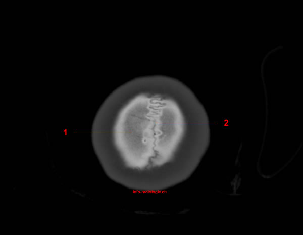
Image 1. CT Anatomy of skull, axial reconstruction, bone window.
1, Parietal bone. 2, Sagittal suture.
-
Image 2. CT Anatomy of skull, axial reconstruction, bone window.
1, Parietal bone. 2, Lambdoid suture. 3, Lambda.
-
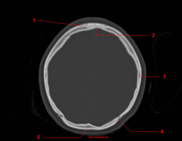
Image 3. CT Anatomy of skull, axial reconstruction, bone window.
1, Sagittal suture. 2, Parietal bone. 3, Lambdoid suture. 4, Occipital bone.
-
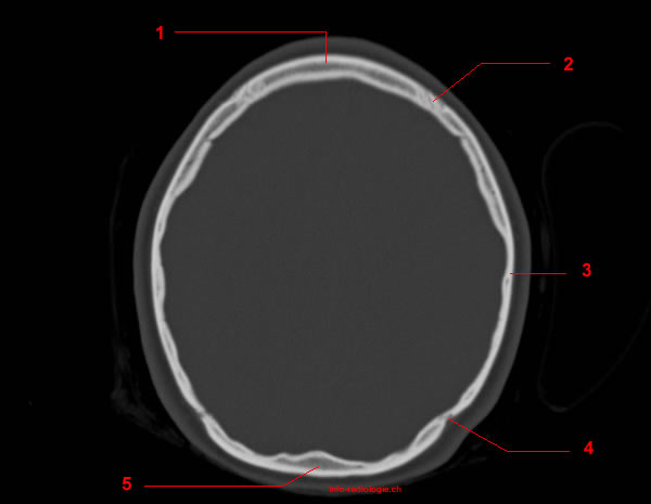
Image 4. CT Anatomy of skull, axial reconstruction, bone window.
1, Coronal suture. 2, Bregma. 3, Parietal bone. 4, Lambdoid suture. 5, Occipital bone.
-
Image 5. CT Anatomy of skull, axial reconstruction, bone window.
1, Frontal bone. 2, Coronal suture. 3, Parietal bone. 4, Lambdoid suture. 5, Occipital bone.
-
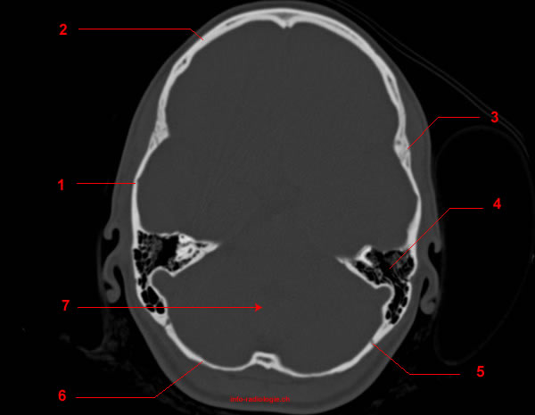
Image 6. CT Anatomy of skull, axial reconstruction, bone window.
1, Temporal bone. 2, Frontal bone. 3, Squamosal suture. 4, Mastoid air cells. 5, Lambdoid suture. 6, Occipital bone. 7, Posterior cranial fossa.
-
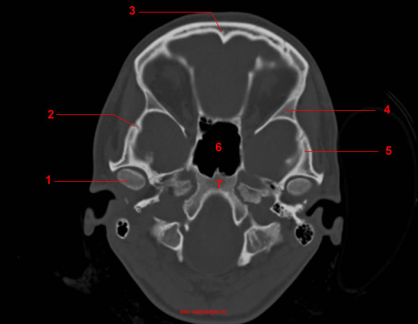
Image 7. CT Anatomy of skull, axial reconstruction, bone window.
1, Middle cranial fossa. 2, Greater wing of sphenoid bone. 3, Frontal bone. 4, Anterior cranial fossa. 5, external auditory canal. 6, Petrous temporal bone.
-
Image 8. CT Anatomy of skull, axial reconstruction, bone window.
1, Mandibular condyle. 2, Sphenosquamosal suture. 3, Frontal bone. 4, Greater wing of sphenoid bone. 5, Temporal bone. 6, Sphenoid sinus. 7, Clivus.
-
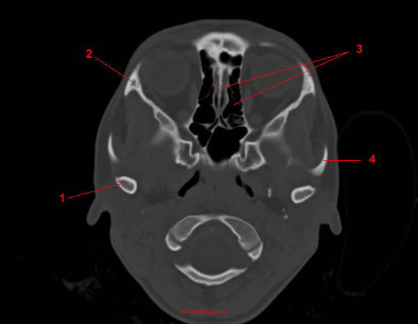
Image 9. CT Anatomy of skull, axial reconstruction, bone window.
1, Mandibular condyle. 2, Zygomatic bone. 3, Ethmoid bone / ethmoid air cells. 4, Zygomatic arch.
-
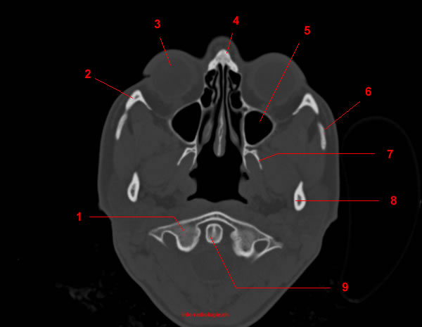
Image 10. CT Anatomy of skull, axial reconstruction, bone window.
1, Lateral mass of C1. 2, Zygomatic bone. 3, Globe. 4, Nasal bone. 5, Maxillary sinus. 6, Zygomatic arch. 7, Pterygoid process (sphenoid bone). 8, Mandible. 9, Ondotoid process (C2).
-
Image 11 of 11. CT Anatomy of skull, axial reconstruction, bone window.
1, C1. 2, Pterygoid process. 3, Maxillary sinus. 4, Zygomatic bone. 5, Mandible. 6, Ondotoid process (C2).