-
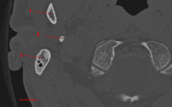
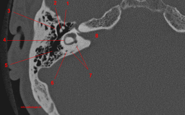
Image 1. CT Anatomy of temporal bone
1, Condylar process of mandible. 2, Styloid process. 3, Mastoid process.
-
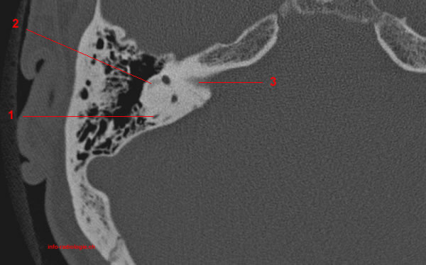
Image 2. CT Anatomy of temporal bone
1, Condylar process of mandible. 2, Canal hypoglosse. 3, Mastoid process.
-
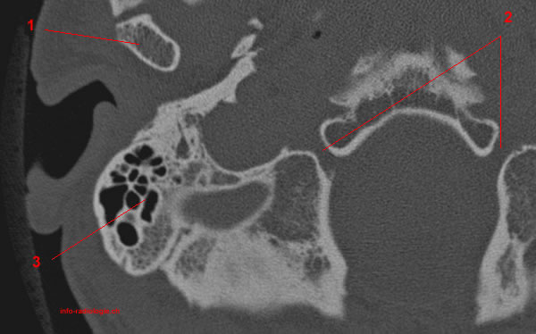
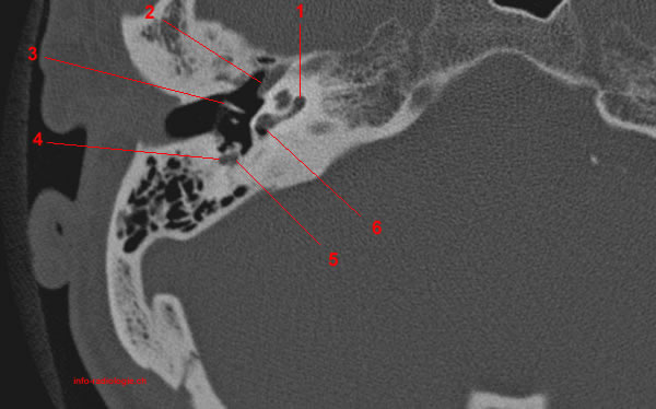
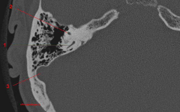
Image 3. CT Anatomy of temporal bone
1, Condylar process of mandible. 2, Carotid canal. 3, Jugular bulb. 4, Facial nerve canal. 5, Sigmoid sinus.
-
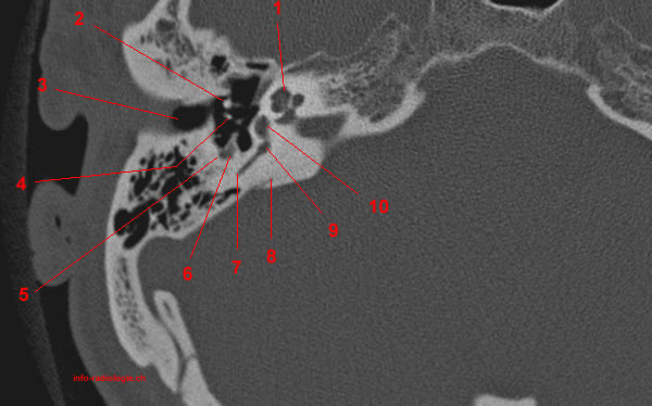
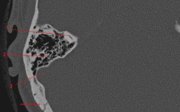
Image 4. CT Anatomy of temporal bone
1, Jugular foramen (pars nervosa). 2, Carotid canal (pars horizontal). 3, Tympan. 4, External auditory canal. 5, Facial nerve canal. 6, Jugular bulb. 7, Sigmoid sinus.
-
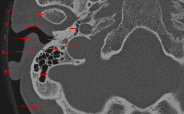
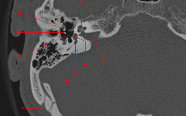
Image 5. CT Anatomy of temporal bone
1, Carotid canal. 2, Tensor tympani muscle. 3, Manubrium of malleus. 4, Facial nerve canal. 5, Sigmoid sinus. 6, Cochlea (basal turn).
-
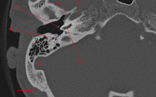
Image 6. CT Anatomy of temporal bone
1, Cochlea (basal turn). 2, Tensor tympani muscle. 3, Manubrium of malleus. 4, Facial nerve canal. 5, Stapedius muscle. 6, Round window.
-
Image 7. CT Anatomy of temporal bone
1, Cochlea. 2, Malleus. 3, External auditory canal. 4, Incus. 5, Facial nerve canal. 6, Stapedius muscle. 7, Posterior semicircular canal. 8, Vestibular aqueduct. 9, Ampulla of posterior semicircular canal. 10, Vestibule.
-
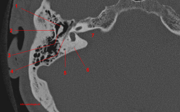
Image 8. CT Anatomy of temporal bone
1, Modiolus. 2, Malleus. 3, Incus. 4, Stapes. 5, Posterior semicircular canal. 6, Ampulla of posterior semicircular canal. 7, Oval window. 8, Internal auditory canal.
-
Image 9. CT Anatomy of temporal bone
1, Malleus. 2, Incus. 3, Facial nerve canal. 4, Posterior semicircular canal. 5, Vestibular aqueduct. 6, Internal auditory canal.
-
Image 10. CT Anatomy of temporal bone
1, Malleus. 2, Koerner septum. 3, Incus. 4, Facial nerve canal. 5, Posterior semicircular canal. 6, Vestibular aqueduct. 7, Internal auditory canal.
-
Image 11. CT Anatomy of temporal bone
1, Incus body. 2, Koerner septum. 3, Malleus head. 4, Geniculate ganglion. 5, Facial nerve canal. 6, Internal auditory canal. 7, Vestibule. 8, Vestibular aqueduct. 9, Posterior semicircular canal.
-
Image 12. CT Anatomy of temporal bone
1, Malleus . 2, Incus. 3, Koerner septum. 4, Lateral semicircular canal. 5, Antrum. 6, Posterior semicircular canal. 7, Vestibular aqueduct. 8, Internal auditory canal.
-
Image 13. CT Anatomy of temporal bone
1, Posterior semicircular canal. 2, Lateral semicircular canal. 3, Internal auditory canal.
-
Image 14. CT Anatomy of temporal bone
1, Posterior semicircular canal. 2, Lateral semicircular canal. 3, Sigmoid sinus.
-
Image 15. CT Anatomy of temporal bone
1, Antrum. 2, tympanic cavity. 3, Superior semicircular canal (anterior part). 4, Petromastoid canal. 5, Superior semicircular canal (posterior part).
-
Image 16 of 16. CT Anatomy of temporal bone
1, Superior semicircular canal. 2, Mastoid air cells. 3, Sigmoid sinus.