CT of the Orbit: anatomy
This webpage presents the anatomical structures found on orbit CT.
Coronal view - Axial view
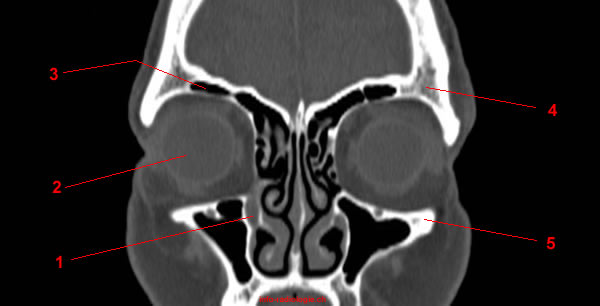
Image 1. CT Anatomy of the orbit. Coronal reconstruction.
1, Nasolacrimal duct. 2, Globe. 3, Frontal sinus. 4, Frontal bone. 5, Zygomatic bone.
-
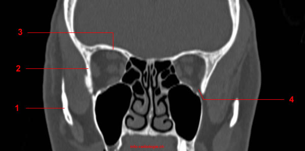
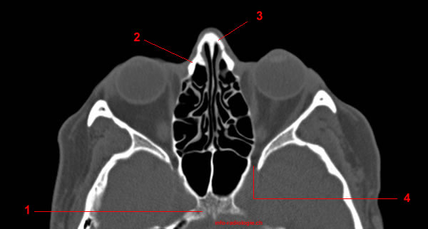
Image 1. CT Anatomy of the orbit. Coronal reconstruction.
1, Nasolacrimal duct. 2, Globe. 3, Frontal sinus. 4, Frontal bone. 5, Zygomatic bone.
-
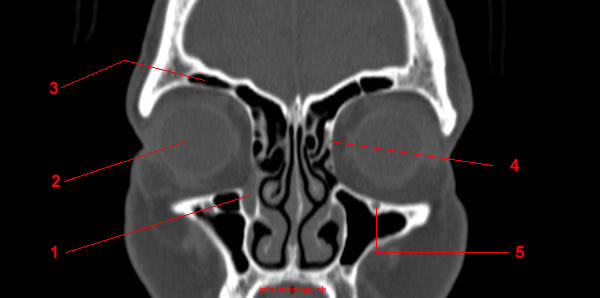
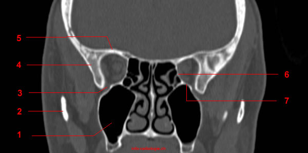
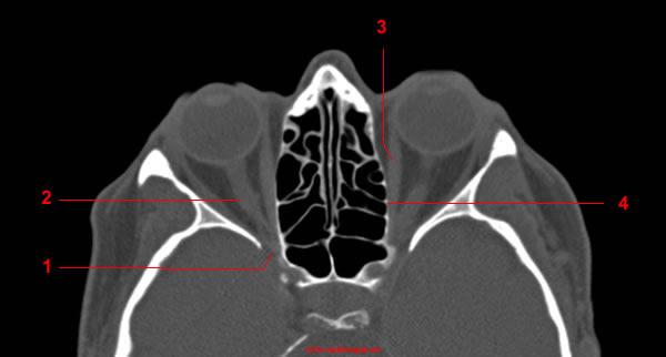
Image 2. CT Anatomy of the orbit. Coronal reconstruction.
1, Nasolacrimal duct. 2, Globe. 3, Frontal sinus. 4, Lamina papyracea of the ethmoid bone. 5, Infraorbital canal.
-
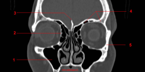
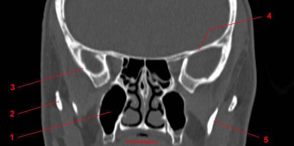
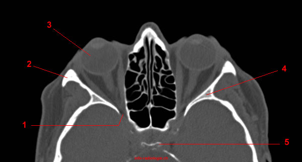
Image 3. CT Anatomy of the orbit. Coronal reconstruction.
1, Maxillary sinus. 2, Lamina papyracea of the ethmoid bone. 3, Crista galli. 4, Frontal bone and sinus. 5, Zygomatic bone.
-
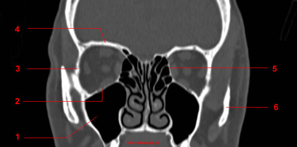
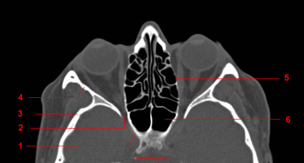
Image 4. CT Anatomy of the orbit. Coronal reconstruction.
1, Maxillary bone and sinus. 2, Lamina papyracea of the ethmoid bone. 3, Cribriform plate (ethmoid). 4, Frontal bone and sinus. 5, Zygomatic bone. 6, Inferior nasal concha.
-
Image 5. CT Anatomy of the orbit. Coronal reconstruction.
1, Maxillary sinus. 2, Maxillary bone. 3, Zygomatic bone. 4, Frontal bone. 5, Lamina papyracea of the ethmoid bone. 6, Zygomatic arch.
-
Image 6. CT Anatomy of the orbit. Coronal reconstruction.
1, Zygomatic arch. 2, Zygomatic bone. 3, Frontal bone.4, Inferior orbital fissure.
-
Image 7. CT Anatomy of the orbit. Coronal reconstruction.
1, Maxillary sinus. 2, Zygomatic arch. 3, Inferior orbital fissure. 4, Zygomatic bone. 5, Frontal bone. 6, Lamina papyracea of the ethmoid bone. 7, Maxillary bone.
-
Image 8. CT Anatomy of the orbit. Coronal reconstruction.
1, Maxillary sinus. 2, Zygomatic arch. 3, Zygomatic bone. 4, Frontal bone. 5, Mandible.
-
Image 9. CT Anatomy of the orbit. Axial reconstruction.
1, Greater wing of sphenoid. 2, Zygomatic bone. 3, Frontal bone.
-
Image 10. CT Anatomy of the orbit. Axial reconstruction.
1, Optic canal. 2, Greater wing of sphenoid. 3, Zygomatic bone. 4, Frontal bone.
-
Image 11. CT Anatomy of the orbit. Axial reconstruction.
1, Sella turcica. 2, Superior orbital fissure. 3, Greater wing of sphenoid. 4, Zygomatic bone. 5, Lamina papyracea of the ethmoid bone. 6, Ethmoidal cells.
-
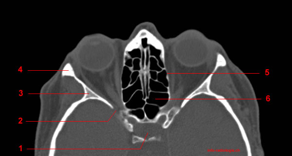
Image 12. CT Anatomy of the orbit. Axial reconstruction.
1, Superior orbital fissure. 2, Optic nerve. 3, Medial rectus muscle. 4, Lamina papyracea of the ethmoid bone.
-
Image 13. CT Anatomy of the orbit. Axial reconstruction.
1, Superior orbital fissure. 2, Zygomatic bone. 3, Globe. 4, Greater wing of sphenoid. 5, Dorsum sellae.
-
Image 14. CT Anatomy of the orbit. Axial reconstruction.
1, sphenoid bone. 2, Superior orbital fissure. 3, Sphenotemporal suture. 4, Sphenozygomatic suture. 5, Lamina papyracea of the ethmoid bone. 6, Sphenoidal sinus.
-
Image 15. CT Anatomy of the orbit. Axial reconstruction.
1, sphenoid bone. 2, Lacrimal bone. 3, Nasal bone. 4, Superior orbital fissure.
-
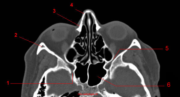
Image 16. CT Anatomy of the orbit. Axial reconstruction.
1, Sphenoidal sinus. 2, Ethmoidal cells. 3, Carotid canal.
-
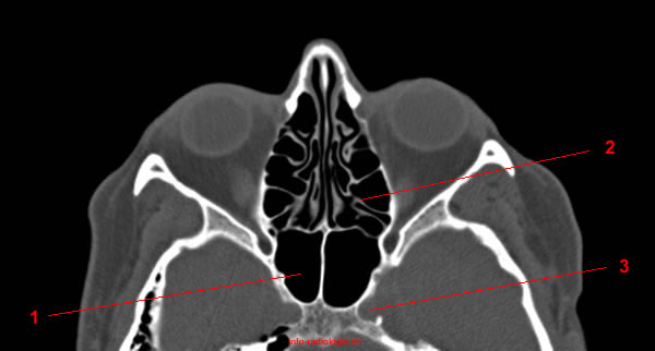
Image 17 of 17. CT Anatomy of the orbit. Axial reconstruction.
1, Foramen rotundem. 2, Zygomatic bone. 3, Maxillary bone. 4, Nasal bone. 5, Inferior orbital fissure. 6, Sphenoidal sinus.
Bony orbit
Lateral wall:
Orbital process of the frontal bone (Anterior superior portion)
Orbital process of the zygomatic bone (Antero inferior portion)
Sphenoid bone (Postero-medial portion)
Medial wall:
Greater wing of the sphenoid (posterior portion)
Ethmoide bone (planum), behind the lacrimal bone
Lacrimal bone (Anterior superior portion, behind the processus frontal of the maxillary bone)
Maxillary bone (Anterior inferior portion)
Sphenoid bone.
Roof:
Orbital process of the frontal bone (anterior superior portion)
Lesser wing of the sphenoid (postero-medial portion)
Inferior wall:
Orbital process of the frontal bone
Orbital process of the zygomatic bone.
Sphenoid bone.
Superior orbital fissure lies between the lesser and the greater wing of sphenoid. This fissure allows the passage to the nerves III, IV, VI, branches of the V(1) and ophthalmic veins.
Inferior orbital fissure lies between the greater wing of sphenoid, the orbital process of the maxillary bone, and, laterally, the zygomatic bone. This fissure allows the passage of branches of the V (2) as well as ophthalmic veins.
Foramen infraorbital (maxillary bone): infraorbital nerve (V2)
Foramen rotundum : V2, proximal segment
Reference:
• Harnsberger HR, Osborn AG, Ross JS, Moore KR, Salzman KL, Carrasco CR, Halmiton BE, Davidson HC, Wiggins RH. Diagnostic and Surgical Imaging Anatomy: Brain, Head and Neck, Spine. 3rd ed. Salt Lake City, Utah. Amirsys. 2007.
• Bourjat P, Veillon F. Imagerie radiologique tête et cou. Paris, Vigot. 1995.
• Gouazé A, Baumann JA, Dhem A. Sobota. Atlas d'Anatomie humaine. Tome 3. Système nerveux central, système nerveux autonome, organe des sens et peau, vaisseaux et nerfs périphériques. 1er éd. Paris, Maloine. 1977.
• Kahle W, Cabrol C. Anatomie. Tome 3: Système nnerveux et organe des sens. 1er éd. Paris, Flammarion. 1979.