Glossopharyngeal nerve
This page describes the path of the glossopharyngeal nerve with brain MRI (axial T1 and T2 weighted images).
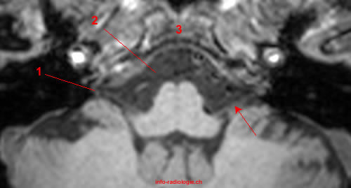
Brain MRI, axial T1-weighted image. Image 1.
1, Jugular foramen. 2, Cerebellomedullary cistern. 3, Clivus. Arrow, Glossopharyngeal nerve (IX).
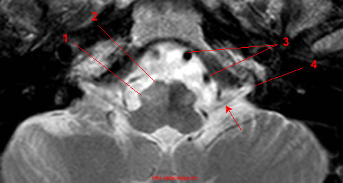
1, Jugular foramen. 2, Cerebellomedullary cistern. 3, Clivus. Arrow, Glossopharyngeal nerve (IX).
Bibliographie:
• Harnsberger HR, Osborn AG, Ross JS, Moore KR, Salzman KL, Carrasco CR, Halmiton BE, Davidson HC, Wiggins RH. Diagnostic and Surgical Imaging Anatomy: Brain, Head and Neck, Spine. 3rd ed. Salt Lake City, Utah. Amirsys. 2007.