-
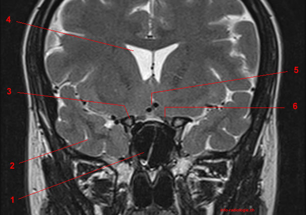
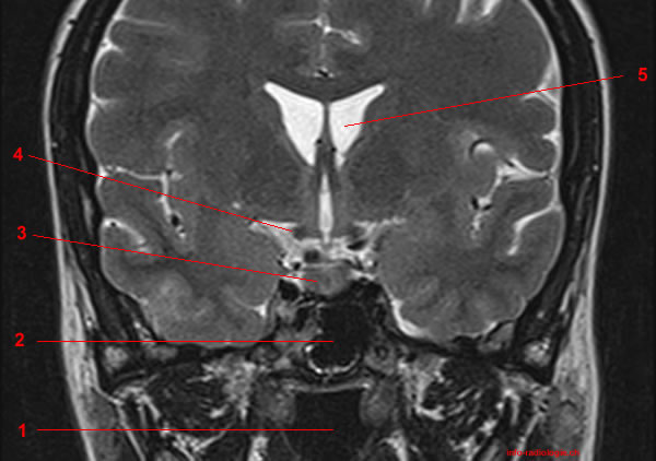
MRI of the pituitary gland: T2-weighted coronal view. Image 1. 1, Sphenoidal sinus 2, Anterior clinoid process. 3, Lateral ventricle. 4, Anterior cerebral artery. 5, Optic nerve.
-
MRI of the pituitary gland: T2-weighted coronal view. Image 2. 1, Sphenoidal sinus 2, Temporal pole. 3, Anterior clinoid process. 4, Lateral ventricle. 5, Anterior cerebral artery. 6, Optic nerve.
-
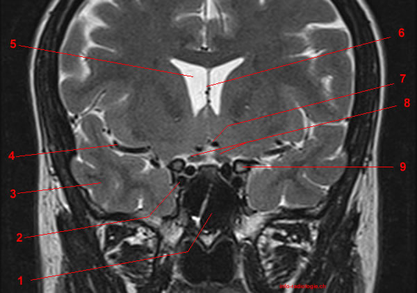
MRI of the pituitary gland: T2-weighted coronal view. Image 3. 1, Sphenoidal sinus 2, Internal carotid artery. 3, Temporal pole. 4, Middle cerebral artery. 5, Lateral ventricle. 6, Septum pellucidum. 7, Anterior cerebral artery. 8, Optic nerve. 9, Anterior clinoid process.
-
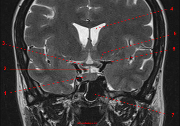
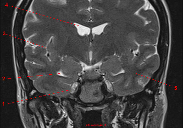
MRI of the pituitary gland: T2-weighted coronal view. Image 4. 1, Nasopharynx. 2, Sphenoidal sinus 3, Internal carotid artery. 4, Optic chiasm. 5, Lateral ventricle. 6, Anterior cerebral artery. 7, Middle cerebral artery. 8, Temporal pole.
-
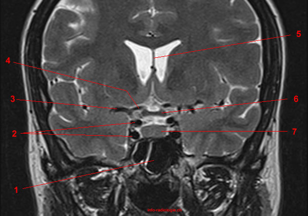
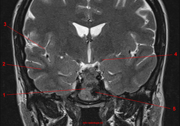
MRI of the pituitary gland: T2-weighted coronal view. Image 5. 1, Sphenoidal sinus 2, Internal carotid artery. 3, Middle cerebral artery. 4, Anterior cerebral artery. 5, Septum pellucidum. 6, Optic chiasm. 7, Pituitary gland.
-
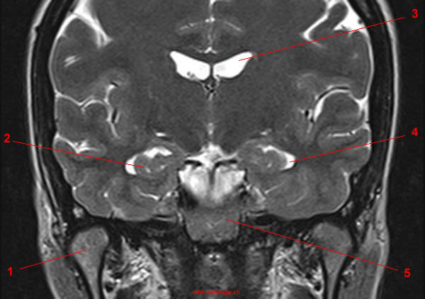
MRI of the pituitary gland: T2-weighted coronal view. Image 6. 1, Pituitary gland. 2, Infudibulum. 3, Chiasma. 4, Lateral ventricle. 5, Anterior cerebral artery. 6, Middle cerebral artery. 7, Sphenoidal sinus.
-
MRI of the pituitary gland: T2-weighted coronal view. Image 7. 1, Nasopharynx. 2, Sphenoidal sinus. 3, Pituitary gland. 4, Optic tract. 5, Lateral ventricle.
-
MRI of the pituitary gland: T2-weighted coronal view. Image 8. 1, Nasopharynx. 2, Pituitary gland. 3, Optic tract. 4, Third ventricle. 5, Lateral ventricle.
-
MRI of the pituitary gland: T2-weighted coronal view. Image 9. 1, Sphenoidal bone. 2, Temporal pole. 3, Lateral fissure (also called Sylvian fissure). 4, Optic tract. 5, Sphenoidal sinus
-
MRI of the pituitary gland: T2-weighted coronal view. Image 10. 1, Meckel's cave (trigeminal cave). 2, Temporal horn. 3, Lateral fissure (also called Sylvian fissure). 4, Lateral ventricle. 5, Temporal pole.
-
MRI of the pituitary gland: T2-weighted coronal view. Image 11. 1, mandible. 2, Lateral ventricle. 3, Temporal horn. 4, Nasopharynx.
-
MRI of the pituitary gland: T2-weighted coronal view. Image 12. 1, mandible. 2, Hippocampus. 3, Lateral ventricle. 4, Temporal horn. 5, Clivus.
-
MRI of the pituitary gland: T1-weighted coronal view .(without iv contrast media) Image 13. 1, Nasopharynx. 2, Sphenoidal sinus 3, Internal carotid artery. 4, Optic chiasm. 5, Lateral ventricle. 6, Anterior cerebral artery. 7, Middle cerebral artery. 8, Temporal pole.
-
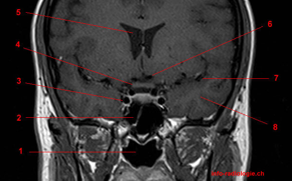
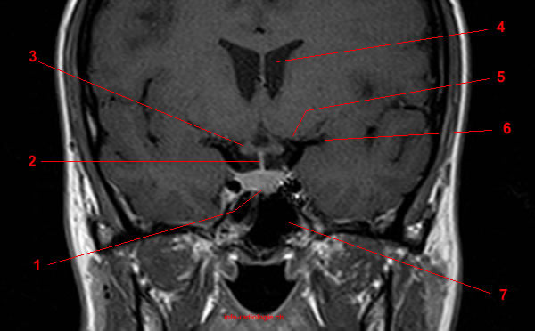
MRI of the pituitary gland: T1-weighted coronal view (after iv contrast media). Image 14. 1, Nasopharynx. 2, Sphenoidal sinus 3, Internal carotid artery. 4, Optic chiasm. 5, Lateral ventricle. 6, Anterior cerebral artery. 7, Middle cerebral artery. 8, Temporal pole.
-
MRI of the pituitary gland: T1-weighted coronal view .(without iv contrast media) Image 15. 1, Sphenoidal sinus 2, Internal carotid artery. 3, Middle cerebral artery. 4, Anterior cerebral artery. 5, Septum pellucidum. 6, Optic chiasm. 7, Pituitary gland.
-
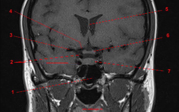
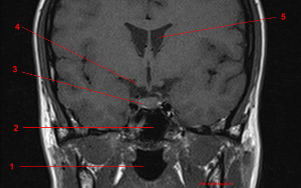
MRI of the pituitary gland: T1-weighted coronal view (after iv contrast media). Image 16. 1, Sphenoidal sinus 2, Internal carotid artery. 3, Middle cerebral artery. 4, Anterior cerebral artery. 5, Septum pellucidum. 6, Optic chiasm. 7, Pituitary gland.
-
MRI of the pituitary gland: T1-weighted coronal view .(without iv contrast media) Image 17. 1, Pituitary gland. 2, Infudibulum. 3, Chiasma. 4, Lateral ventricle. 5, Anterior cerebral artery. 6, Middle cerebral artery. 7, Sphenoidal sinus.
-
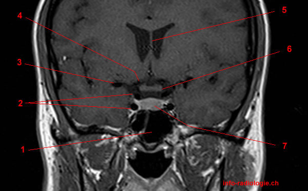
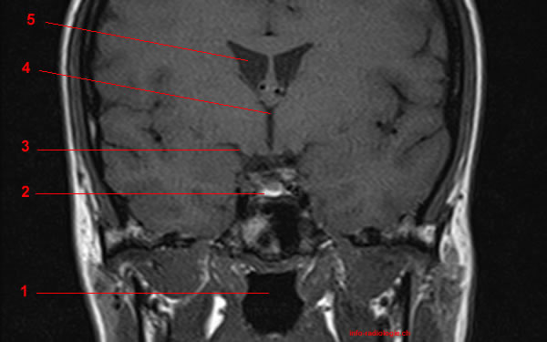
MRI of the pituitary gland: T1-weighted coronal view (after iv contrast media). Image 18. 1, Pituitary gland. 2, Infudibulum. 3, Chiasma. 4, Lateral ventricle. 5, Anterior cerebral artery. 6, Middle cerebral artery. 7, Sphenoidal sinus
-
MRI of the pituitary gland: T1-weighted coronal view .(without iv contrast media) Image 19. 1, Nasopharynx. 2, Sphenoidal sinus. 3, Pituitary gland. 4, Optic tract. 5, Lateral ventricle.
-
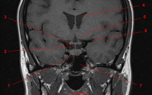
MRI of the pituitary gland: T1-weighted coronal view (after iv contrast media). Image 20. 1, Nasopharynx. 2, Sphenoidal sinus. 3, Pituitary gland. 4, Optic tract. 5, Lateral ventricle.
-
MRI of the pituitary gland: T1-weighted coronal view .(without iv contrast media) Image 21. 1, Nasopharynx. 2, Pituitary gland. 3, Optic tract. 4, Third ventricle. 5, Lateral ventricle.
-
MRI of the pituitary gland: T1-weighted coronal view (after iv contrast media). Image 22. 1, Nasopharynx. 2, Pituitary gland. 3, Optic tract. 4, Third ventricle. 5, Lateral ventricle.
-
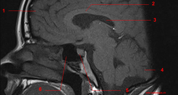
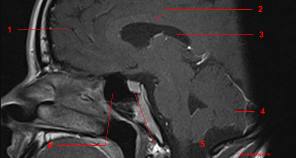
MRI of the pituitary gland: T1-weighted sagittal view .(without iv contrast media) Image 23. 1, Frontal pole. 2, Corpus callosum. 3, Lateral ventricle. 4, Cerebellum. 5, Pituitary gland. 6, Sphenoidal sinus
-
MRI of the pituitary gland: T1-weighted coronal view (after iv contrast media). Image 24. 1, Frontal pole. 2, Corpus callosum. 3, Lateral ventricle. 4, Cerebellum. 5, Pituitary gland. 6, Sphenoidal sinus
-
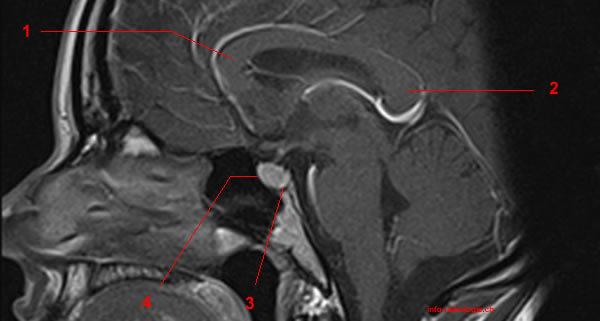
MRI of the pituitary gland: T1-weighted sagittal view (without iv contrast media). Image 25. 1, Genu of corpus callosum. 2, Splenium of corpus callosum. 3, Neurohypophysis. 4, Antehypophysis.
-
MRI of the pituitary gland: T1-weighted coronal view (after iv contrast media). Image 26. 1, Genu of corpus callosum. 2, Splenium of corpus callosum. 3, Neurohypophysis. 4, Antehypophysis.
-
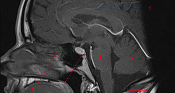
MRI of the pituitary gland: T1-weighted sagittal view (without iv contrast media). Image 27. 1, Lateral ventricle. 2, Cerebellum. 3, Pons. 4, Neurohypophysis. 5, Antehypophysis.
-
MRI of the pituitary gland: T1-weighted coronal view (after iv contrast media). Image 28. 1, Lateral ventricle. 2, Cerebellum. 3, Pons. 4, Neurohypophysis. 5, Antehypophysis.
-
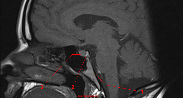
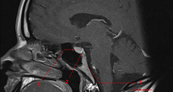
MRI of the pituitary gland: T1-weighted sagittal view (without iv contrast media). Image 29. 1, Clivus. 2, Neurohypophysis. 3, Antehypophysis.
-
MRI of the pituitary gland: T1-weighted coronal view (after iv contrast media). Image 30 of 30. 1, Clivus. 2, Neurohypophysis. 3, Antehypophysis.
