-
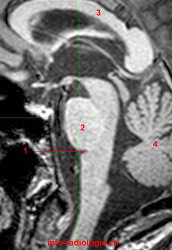
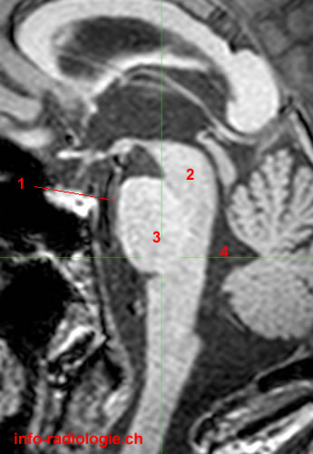
MRI of the brain, T1-weighted sagittal view. Image 1. 1, Pontomedullary sulcus. 2, Pons. 3, Corpus callosum. 4, Cerebellum.
-
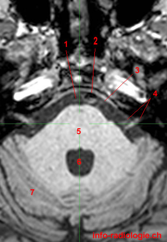
MRI of the brain, T1-weighted axial view. Image 2. 1, Prepontine cistern. 2, Basilar artery. 3, Pons. 4, Fourth ventricle. 5, Central lobule. 6, Culmen.
-
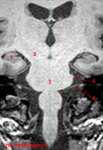
MRI of the brain, T1-weighted coronal cut. Image 3. 1, Lateral ventricle. 2, Third ventricle. 3, Interpeduncular cistern. 4, Pons. 5, Trigeminal nerve (V).
-
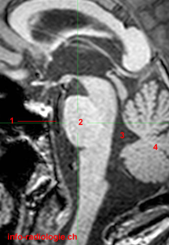
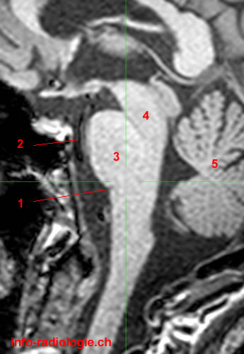
MRI of the brain, T1-weighted sagittal cut. Image 4. 1, Basilar artery. 2, Pons. 3, Fourth ventricle. 4, Cerebellum.
-
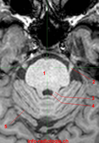
MRI of the brain, T1-weighted axial view. Image 5. 1, Pons. 2, Trigeminal nerve (V). 3, Superior cerebellar peduncle. 4, Fourth ventricle. 5, Quadrangular lobule.
-
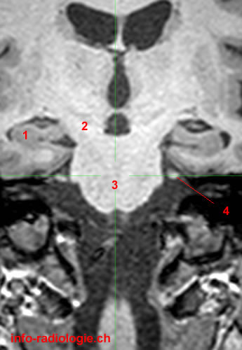
MRI of the brain, T1-weighted coronal view. Image 6. 1, Hippocampus. 2, Cerebral peduncle. 3, Pons. 4, Trigeminal nerve (V).
-
MRI of the brain, T1-weighted sagittal view. Image 7. 1, Prepontine cistern. 2, Midbrain. 3, Pons. 4, Fourth ventricle.
-
MRI of the brain, T1-weighted axial view. Image 8. 1, Prepontine cistern. 2, Basilar artery. 3, Abducens nerve (VI). 4, Vestibulocochlear nerve (VII) and Facial nerve (VII). 5, Pons. 6, Fourth ventricle. 7, Cerebellar hemisphere.
-
MRI of the brain, T1-weighted coronal view. Image 9. 1, Hippocampus. 2, Cerebral peduncle. 3, Pons. 4, Vestibulocochlear nerve (VII) and Facial nerve (VII). 5, Middle cerrebelar peduncle.
-
MRI of the brain, T1-weighted sagittal view. Image 10. 1, Pontomedullary sulcus. 2, Basilar artery. 3, Pons. 4, Midbrain. 5, Cerebellum.
-
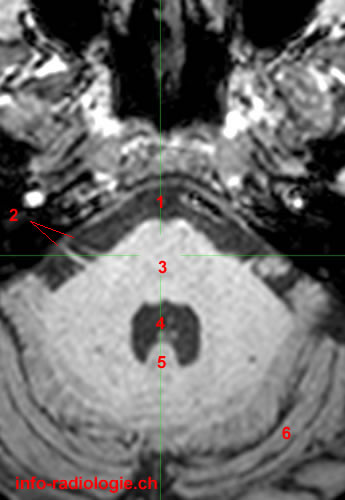
MRI of the brain, T1-weighted axial view. Image 11. 1, Prepontine cistern. 2, Vestibulocochlear nerve (VII) and Facial nerve (VII). 3, Pons. 4, Fourth ventricle. 5, Nodulus (Vermis). 6, Cerebellar hemisphere.
-
MRI of the brain, T1-weighted coronal view. Image 12. 1, Hippocampus. 2, Third ventricle. 3, Midbrain. 4, Pons. 5, Pontomedullary sulcus. 6, Vestibulocochlear nerve (VII) and Facial nerve (VII).