-
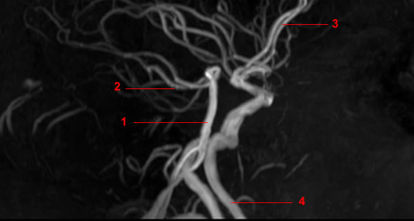
MRA of Circle of Willis. lateral view with rotations. Image 1. 1, Basilar artery. 2, Posterior cerebral artery. 3, Anterior cerebral artery (A2). 4, Internal carotid artery.
-
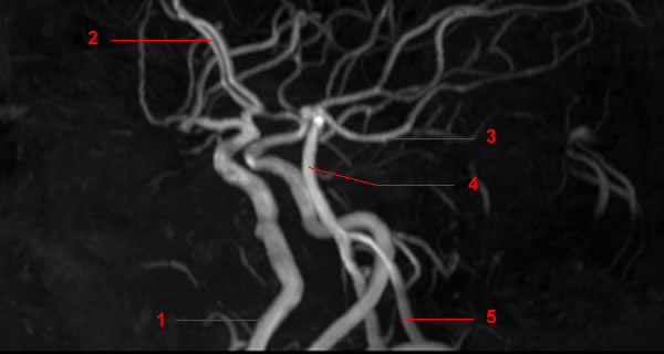
MRA of Circle of Willis. lateral view with rotations. Image 2. 1, Vertebral artery. 2, Basilar artery. 3, Anterior cerebral artery. 4, Internal carotid artery.
-
MRA of Circle of Willis. lateral view with rotations. Image 3. 1, Vertebral artery. 2, Internal carotid artery. 3, Basilar artery. 4, Anterior cerebral artery. 5, Middle cerebral artery.
-
MRA of Circle of Willis. lateral view with rotations. Image 4. 1, Vertebral artery. 2, Internal carotid artery. 3, Basilar artery. 4, Anterior cerebral artery. 5, Middle cerebral artery.
-
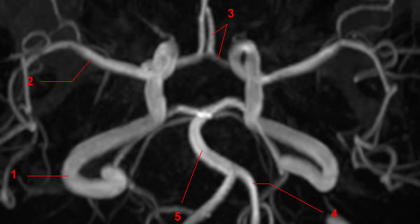
MRA of Circle of Willis. lateral view with rotations. Image 5. 1, Internal carotid artery. 2, Vertebral artery. 3, Basilar artery. 4, Middle cerebral artery. 5, Anterior cerebral artery.
-
MRA of Circle of Willis. lateral view with rotations. Image 6. 1, Internal carotid artery. 2, Vertebral artery. 3, Basilar artery. 4, Middle cerebral artery. 5, Anterior cerebral artery.
-
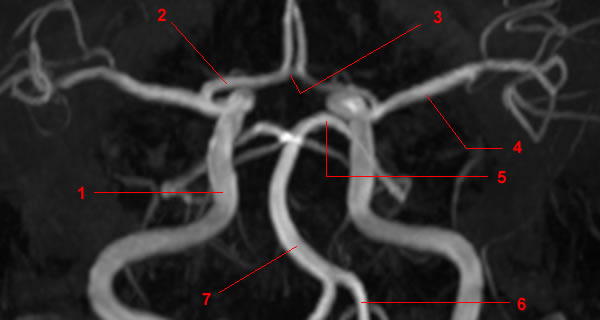
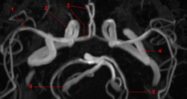
MRA of Circle of Willis. lateral view with rotations. Image 7. 1, Internal carotid artery. 2, Vertebral artery. 3, Basilar artery. 4, Middle cerebral artery. 5, Anterior cerebral artery. 6, Posterior cerebral artery.
-
MRA of Circle of Willis. lateral view with rotations. Image 8. 1, Internal carotid artery. 2, Vertebral artery. 3, Basilar artery. 4, Middle cerebral artery. 5, Anterior cerebral artery. 6, Posterior cerebral artery.
-
MRA of Circle of Willis. lateral view with rotations. Image 9. 1, Internal carotid artery. 2, Vertebral artery. 3, Basilar artery. 4, Middle cerebral artery. 5, Anterior cerebral artery. 6, Posterior cerebral artery.
-
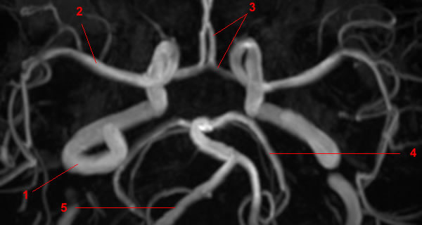
MRA of Circle of Willis. lateral view with rotations. Image 10. 1, Vertebral artery. 2, Internal carotid artery. 3, Middle cerebral artery. 4, Anterior cerebral artery. 5, Posterior cerebral artery. 6, Basilar artery.
-
MRA of Circle of Willis. lateral view with rotations. Image 11. 1, Internal carotid artery. 2, Anterior cerebral artery. 3, Posterior cerebral artery. 4, Basilar artery. 5, Vertebral artery.
-
MRA of Circle of Willis. lateral view with rotations. Image 12 of 12. 1, Internal carotid artery. 2, Anterior cerebral artery. 3, Posterior cerebral artery. 4, Basilar artery. 5, Vertebral artery.
