Hypothalamus
The hypothalamus is a portion of the brain near the third ventricle, located below the thalamus and above the brainstem. The anterior boundary of the hypothalamus is determined by the line connecting the anterior commissure, the lamina terminalis and the optic chiasm. The lower limit of the hypothalamus is formed by the infundibulum, the tuber cinerum and the mamillary bodies. The posterior limit is represented by a straight line joining the mamillary bodies and the posterior commissure.
The hypothalamus is involved in the following control systems:
• body temperature
• autonomic nervous system
• emotional and food behavior
• endocrine (via the pituitary)
• circadian rhythm.
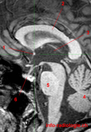
MRI of the Brain (hypothalamus): T1-weighted sagittal cut. Image 1.
1, Anterior commissure. 2, Corpus callosum. 3, Third ventricle. 4, Cerebellum. 5, Pons. 6, Pituitary gland.
-
MRI of the Brain (hypothalamus): T1-weighted sagittal cut. Image 1.
1, Anterior commissure. 2, Corpus callosum. 3, Third ventricle. 4, Cerebellum. 5, Pons. 6, Pituitary gland.
-
MRI of the Brain (hypothalamus): T1-weighted axial cut. Image 2.
1, Anterior commissure. 2, Putamen. 3, Third ventricle. 4, Corpus callosum.
-
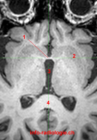
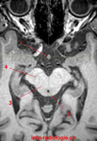
MRI of the Brain (hypothalamus): T1-weighted coronal cut. Image 3.
1, Anterior commissure. 2, Caudate nucleus. 3, Corpus callosum. 4, Lateral ventricle. 5, Third ventricle.
-
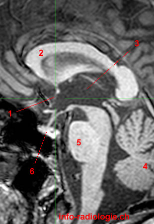
MRI of the Brain (hypothalamus): T1-weighted sagittal cut. Image 4.
1, Lamina terminalis. 2, Corpus callosum. 3, Third ventricle. 4, Cerebellum. 5, Pons. 6, Pituitary gland.
-
MRI of the Brain (hypothalamus): T1-weighted axial cut. Image 5.
1, Lamina terminalis. 2, Third ventricle. 3, Pineal gland. 4, Corpus callosum.
-
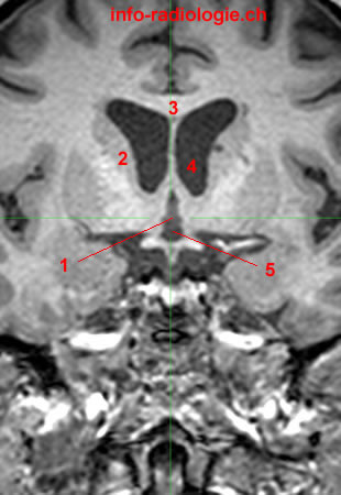
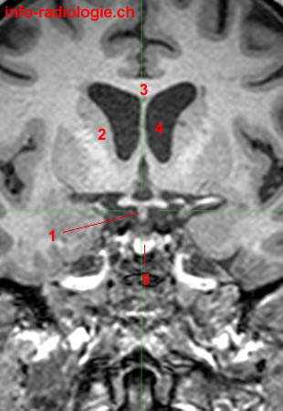
MRI of the Brain (hypothalamus): T1-weighted coronal cut. Image 6.
1, Lamina terminalis. 2, Caudate nucleus. 3, Corpus callosum. 4, Lateral ventricle. 5, Third ventricle.
-
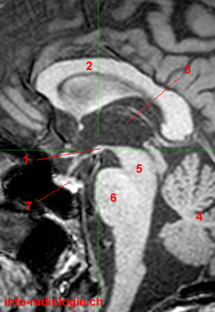
MRI of the Brain (hypothalamus): T1-weighted sagittal cut. Image 7.
1, Optic chiasm. 2, Corpus callosum. 3, Third ventricle. 4, Cerebellum. 5, Pons. 6, Pituitary gland.
-
MRI of the Brain (hypothalamus): T1-weighted axial cut. Image 8.
1, Optic chiasm. 2, Cerebral aqueduct. 3, Superior colliculus.
-
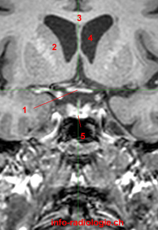
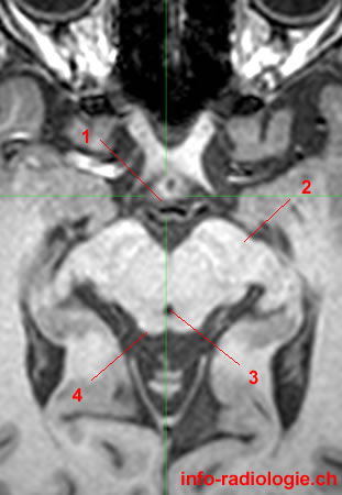
MRI of the Brain (hypothalamus): T1-weighted coronal cut. Image 9.
1, Optic chiasm. 2, Caudate nucleus. 3, Corpus callosum. 4, Lateral ventricle. 5, Pituitary gland.
-
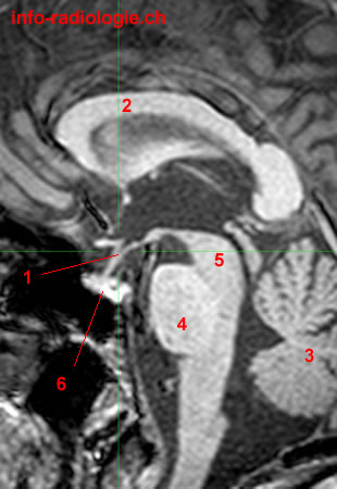
MRI of the Brain (hypothalamus): T1-weighted sagittal cut. Image 10.
1, Infudibulum.
2, Corpus callosum.
3, Cerebellum.
4, Pons.
5, Midbrain.
6, Pituitary gland.
-
MRI of the Brain (hypothalamus): T1-weighted axial cut. Image 11.
1, Infudibulum.
2, Cerebral aqueduct.
3, Midbrain.
4, Substantia nigra.
-
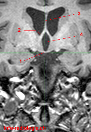
MRI of the Brain (hypothalamus): T1-weighted coronal cut. Image 12.
1, Infudibulum.
2, Caudate nucleus.
3, Corpus callosum.
4, Lateral ventricle.
5, Pituitary gland.
-
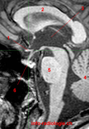
MRI of the Brain (hypothalamus): T1-weighted sagittal cut. Image 13.
1, Tuber cinerum.
2, Corpus callosum.
3, Fornix.
4, Third ventricle.
5, Cerebellum.
6, Fourth ventricle.
7, Pons.
-
MRI of the Brain (hypothalamus): T1-weighted axial cut. Image 14.
1, Tuber cinerum.
2, Cerebral peduncle.
3, Cerebral aqueduct.
4, Superior colliculus.
-
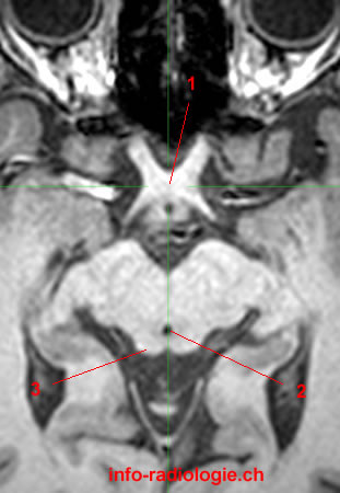
MRI of the Brain (hypothalamus): T1-weighted coronal cut. Image 15.
1, Tuber cinerum. 2, Column of fornix. 3, Septum pellucidum. 4, Third ventricle.
-
MRI of the Brain (hypothalamus): T1-weighted sagittal cut. Image 16.
1, Mammillary body. 2, Corpus callosum. 3, Third ventricle. 4, Cerebellum. 5, Midbrain. 6, Pons. 7, Pituitary gland.
-
MRI of the Brain (hypothalamus): T1-weighted axial cut. Image 17.
1, Mammillary body. 2, Cerebral aqueduct. 3, Superior colliculus.
-
MRI of the Brain (hypothalamus): T1-weighted coronal cut. Image 18.
1, Mammillary body. 2, Corpus callosum. 3, Lateral ventricle. 4, Third ventricle.
-
MRI of the Brain (hypothalamus): T1-weighted sagittal cut. Image 19.
1, Posterior commissure. 2, Midbrain. 3, Pituitary gland. 4, Récessus optique. 5, Corpus callosum.
-
MRI of the Brain (hypothalamus): T1-weighted axial cut. Image 20.
1, Posterior commissure. 2, Third ventricle. 3, Globe (left side).
-
MRI of the Brain (hypothalamus): T1-weighted coronal cut. Image 21 of 21.
1, Posterior commissure. 2, Thalamus. 3, Lateral ventricle. 4, Cerebral aqueduct.