Cerebellum
This photo gallery presents the anatomy of Cerebellum by means of MRI (T1-weighted sagittal, axial and coronal views).
MRI of the brain, T1-weighted axial view. Level: Medulla. Image 1.
1, Cerebellar hemisphere. 2, Medulla. 3, Cerebellar tonsil.
-
MRI of the brain, T1-weighted axial view. Level: Medulla. Image 1.
1, Cerebellar hemisphere. 2, Medulla. 3, Cerebellar tonsil.
-
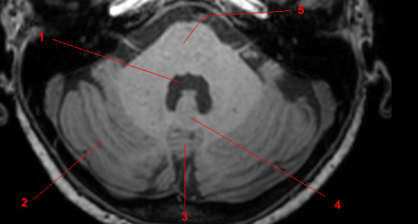
MRI of the brain, T1-weighted axial view. Level: bulbe rachidien. Image 2.
1, Flocculus. 2, Cerebellar tonsil. 3, Vermis. 4, Cerebellar hemisphere. 5, Inferior cerebellar peduncle.
-
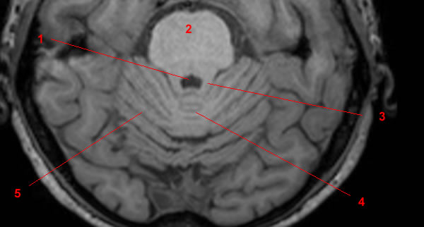
MRI of the brain, T1-weighted axial view. Level: Pons. Image 3.
1, Fourth ventricle. 2, Cerebellar hemisphere. 3, Vermis. 4, Nodulus. 5, Pons.
-
MRI of the brain, T1-weighted axial view. Level: Pons. Image 4.
1, Fourth ventricle. 2, Cerebellar hemisphere. 3, Middle cerebellar peduncle. 4, Pons.
-
MRI of the brain, T1-weighted axial view. Level: pons (superior). Image 5.
1, Fourth ventricle. 2, Pons. 3, Superior cerebellar peduncle. 4, Vermis. 5, Cerebellar hemisphere.
-
MRI of the brain, T1-weighted axial view. Level: Midbrain. Image 6.
1, Midbrain. 2, Vermis.
-
MRI of the brain, T1-weighted coronal view. Image 7.
1, Temporal pole (right side). 2, Middle cerebellar peduncle. 3, Flocculus. 4, Cerebellar hemisphere.
-
MRI of the brain, T1-weighted coronal view. Image 8.
1, Superior cerebellar peduncle. 2, Tentorium cerebelli. 3, Fourth ventricle. 4, Cerebellar hemisphere.
-
MRI of the brain, T1-weighted coronal view. Image 9.
1, Temporal pole (right side). 2, Fourth ventricle. 3, Superior cerebellar peduncle. 4, Horizontal fissure.
-
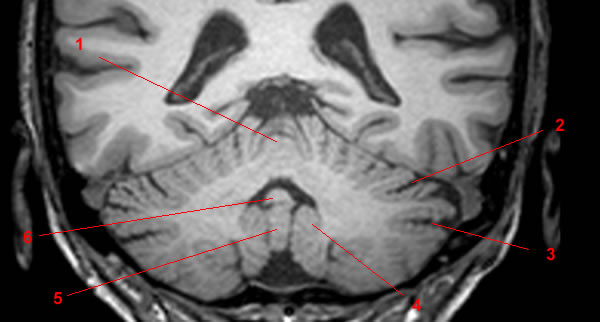
MRI of the brain, T1-weighted coronal view. Image 10.
1, Lobule central. 2, Primary fissure. 3, Horizontal fissure. 4, Cerebellar tonsil. 5, Uvula of vermis. 6, Nodule of vermis.
-
MRI of the brain, T1-weighted coronal view. Image 11.
1, Vermis. 2, Primary fissure. 3, Horizontal fissure.
-
MRI of the brain, T1-weighted sagittal view Image 12.
1, Tentorium cerebelli. 2, Superior cerebellar hemisphere. 3, Horizontal fissure. 4, Horizontal fissure. 5, Cerebellar tonsil.
-
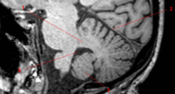
MRI of the brain, T1-weighted sagittal view Image 13.
1, Middle cerebellar peduncle. 2, Tentorium cerebelli. 3, Cerebellar tonsil.
-
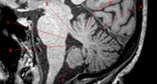
MRI of the brain, T1-weighted sagittal view Image 14.
1, Fourth ventricle. 2, Superior cerebellar peduncle. 3, Culmen. 4, Primary fissure. 5, Cerebellar tonsil. 6, Inferior medullary velum.
-
MRI of the brain, T1-weighted sagittal view Image 15 of 15.
1, Fourth ventricle. 2, Superior medullary velum. 3, Quadrigeminal plate. 4, Culmen. 5, Primary fissure. 6, Declive. 7, Tuber. 8, Pyramide 9, Nodulus (racine) 10, Cerebellar tonsil.
Reference
• Harnsberger HR, Osborn AG, Ross JS, Moore KR, Salzman KL, Carrasco CR, Halmiton BE, Davidson HC, Wiggins RH. Diagnostic and Surgical Imaging Anatomy: Brain, Head and Neck, Spine. 3rd ed. Salt Lake City, Utah. Amirsys. 2007.
