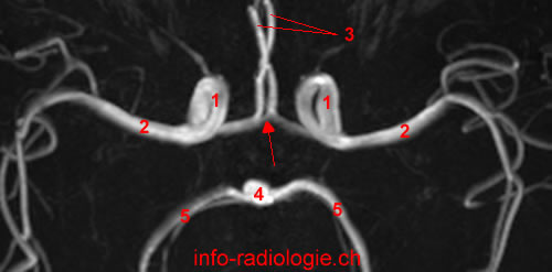
The Internal carotid artery divides into middle cerebral artery and anterior cerebral artery. The anterior cerebral artery enters the longitudinal interhemispheric fissure of the brain.
The anterior communicating artery connects right anterior communicating artery to left anterior communicating artery.
The anterior cerebral artery is divided into 3 parts:
A1 segment, horizontal, get around in the region of the optic nerve.
A2 segment, vertical, means the branch located in the interhemispheric fissure, up before the rostrum of the corpus callosum
A3 segment, distal, courses around the genu of corpus callosum. This segment A3 divides into pericallosal artery and callosomarginal artery.
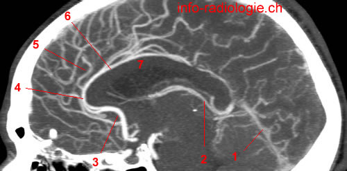
The anterior cerebral arteries irrigate:
• a large part of the medial region of the cerebral hemispheres (2/3 anterior)
• genu of corpus callosum
• anterior limb of the internal capsule
• head of nucleus caudatus.

• Dimmick SJ, Faulder KC. Normal variants of the cerebral circulation at multidetector CT angiography. Radiographics. 2009 Jul-Aug;29(4):1027-43.
• Harnsberger HR, Osborn AG, Ross JS, Moore KR, Salzman KL, Carrasco CR, Halmiton BE, Davidson HC, Wiggins RH. Diagnostic and Surgical Imaging Anatomy: Brain, Head and Neck, Spine. 3rd ed. Salt Lake City, Utah. Amirsys. 2007.
• Bourjat P, Veillon F. Imagerie radiologique tête et cou. Paris, Vigot. 1995.
• Gouazé A, Baumann JA, Dhem A. Sobota. Atlas d'Anatomie humaine. Tome 3. Système nerveux central, système nerveux autonome, organe des sens et peau, vaisseaux et nerfs périphériques. 1er éd. Paris, Maloine. 1977.
• Kahle W, Cabrol C. Anatomie. Tome 3: Système nnerveux et organe des sens. 1er éd. Paris, Flammarion. 1979.