-
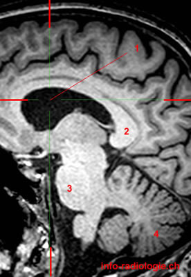
Level 1. Image 1. MRI of brain (magnification), sagittal T1-weighted image. 1, Lateral ventricle. 2, Splenium of corpus callosum. 3, Pons. 4, Cerebellum.
-
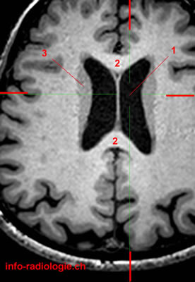
Level 1. Image 2. MRI of brain (magnification), axial T1-weighted image. 1, Lateral ventricle. 2, Corpus callosum. 3, Caudatus nucleus.
-
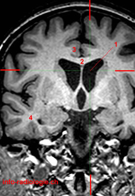
Level 1. Image 3. MRI of brain (magnification), coronal T1-weighted image. 1, Lateral ventricle. 2, Corpus callosum. 3, Cingulate gyrus. 4, Temporal lobe.
-
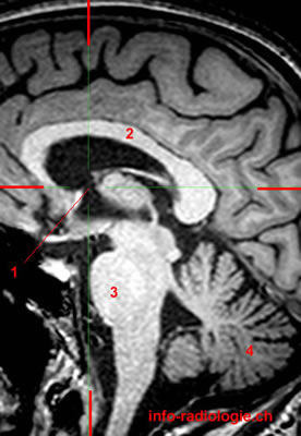
Level 2. Image 1. MRI of brain (magnification), sagittal T1-weighted image. 1, Interventricular foramen (Foramen of Monro) . 2, Corpus callosum. 3, Pons. 4, Cerebellum.
-
Level 2. Image 2. MRI of brain (magnification), axial T1-weighted image. 1, Interventricular foramen (Foramen of Monro) . 2, Thalamus. 3, Lenticular nucleus. 4, Caudatus nucleus.
-
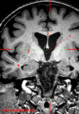
Level 2. Image 3. MRI of brain (magnification), coronal T1-weighted image. 1, Interventricular foramen (Foramen of Monro) . 2, Corpus callosum. 3, Lenticular nucleus. 4, Temporal lobe.
-
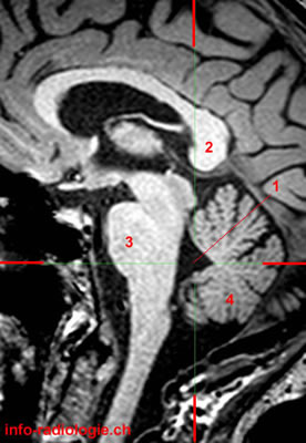
Level 3. Image 1. MRI of brain (magnification), sagittal T1-weighted image. 1, Third ventricle. 2, Fornix. 3, Splenium of corpus callosum. 4, Pons. 5, Cerebellum.
-
Level 3. Image 2. MRI of brain (magnification), axial T1-weighted image. 1, Third ventricle. 2, Thalamus. 3, Lenticular nucleus. 4, Caudatus nucleus.
-
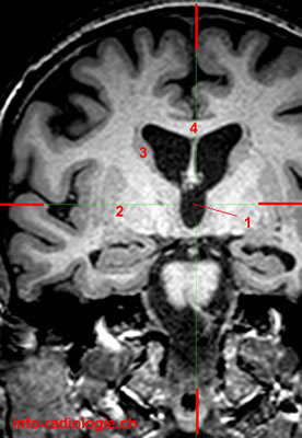
Level 3. Image 3. MRI of brain (magnification), coronal T1-weighted image. 1, Third ventricle. 2, Lenticular nucleus. 3, Caudatus nucleus. 4, Corpus callosum.
-
Level 4. Image 1. MRI of brain (magnification), sagittal T1-weighted image. 1, Cerebral aqueduct. 2, Corpus callosum. 3, Pons. 4, Cerebellum.
-
Level 4. Image 2. MRI of brain (magnification), axial T1-weighted image. 1, Cerebral aqueduct. 2, Temporal lobe (Right side). 3, Globe.
-
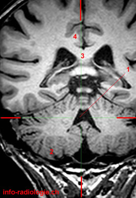
Level 4. Image 3. MRI of brain (magnification), coronal T1-weighted image. 1, Cerebral aqueduct. 2, Cerebellum. 3, Temporal lobe (right side). 4, Corpus callosum.
-
Level 5 (last level). Image 1. MRI of brain (magnification), sagittal T1-weighted image. 1, Fourth ventricle. 2, Splenium of corpus callosum. 3, Pons. 4, Cerebellum.
-
Level 5 (last level). Image 2. MRI of brain (magnification), axial T1-weighted image. 1, Fourth ventricle. 2, Cerebellar hemisphere. A, Anterior. P, Posterior.
-
Level 5 (last level). Image 3. MRI of brain (magnification), coronal T1-weighted image. 1, Fourth ventricle. 2, Cerebellum. 3, Corpus callosum. 4, Cingulate gyrus.