-
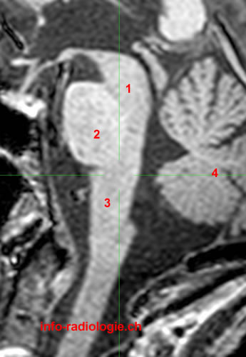
MRI of the brain, T1-weighted sagittal view. Image 1. 1, Midbrain. 2, Pons. 3, Medulla. 4, Cerebellum.
-
MRI of the brain, T1-weighted axial view. Image 2. 1, Flocculus (Cerebellum). 2, Ventral median fissure. 3, Medulla. 4, Inferior cerebellar peduncle. 5, Cerebellar hemisphere. 6, Nodulus (Vermis).
-
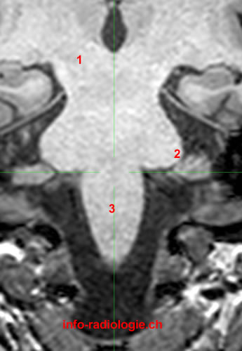
MRI of the brain, T1-weighted coronal view. Image 3. 1, Cerebral peduncle. 2, Middle cerebellar peduncle. 3, Medulla.
-
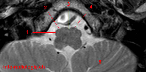
MRI of the brain, T1-weighted axial view. Image 4. 1, Tonsil (Cerebellum). 2, Glossopharyngeal nerve (IX) and vagus nerve (X). 3, Medulla. 4, Basilar artery. 5, Cerebellar hemisphere.
-
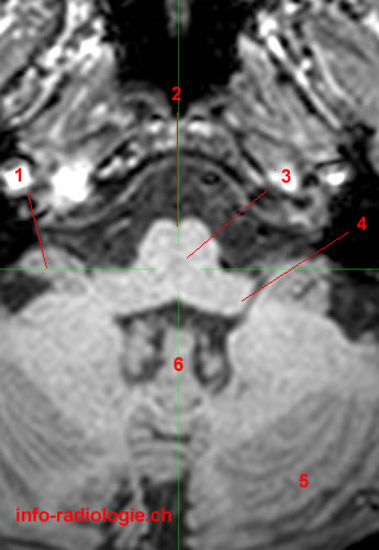
MRI of the brain, T2-weighted axial view. Image 5. 1, Medullary olive. 2, Pre-olivary sulcus. 3, Ventral median fissure. 4, Medullary pyramid. 5, Cerebellar hemisphere.
-
MRI of the brain, T2-weighted axial view. Image 6 of 6. 1, Glossopharyngeal nerve (IX) and vagus nerve (X). 2, Medulla. 3, Premedullary cistern. 4, Jugular foramen. 5, Cerebellar hemisphere.
