Temporal Lobe
The temporal lobe is located in the lower part of the cerebral hemispheres:
• Inferior to the frontal lobe and parietal lobe.
The temporal lobe is separated from the frontal lobe by the lateral sulcus (or Sylvian fissure).
• Anterior to the occipital lobe.
The temporal lobe is virtually continuous with the occipital lobe.
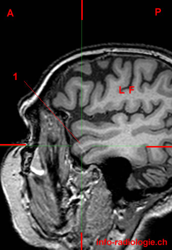
MRI of the brain, T1-weighted sagittal view. Level 1. Image 1.
1, Superior temporal gyrus. LF, Frontal lobe. A, Anterior P, Posterior
-
MRI of the brain, T1-weighted sagittal view. Level 1. Image 1.
1, Superior temporal gyrus. LF, Frontal lobe. A, Anterior P, Posterior
-
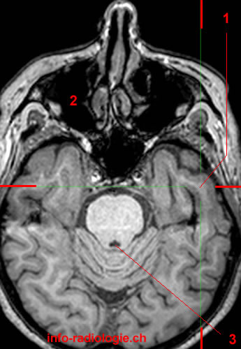
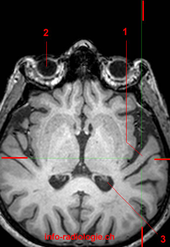
MRI of the brain, T1-weighted axial view. Level 1. Image 2.
1, Superior temporal gyrus. 2, Globe (left side). 3, Lateral ventricle.
-
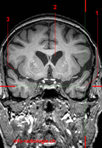
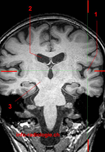
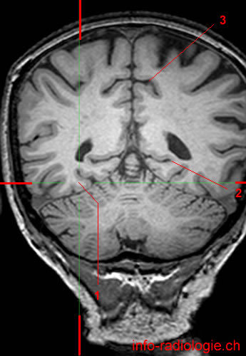
MRI of the brain, T1-weighted coronal view. Level 1. Image 3.
1, Superior temporal gyrus. 2, Corpus callosum. 3, Superior frontal gyrus (right side).
-
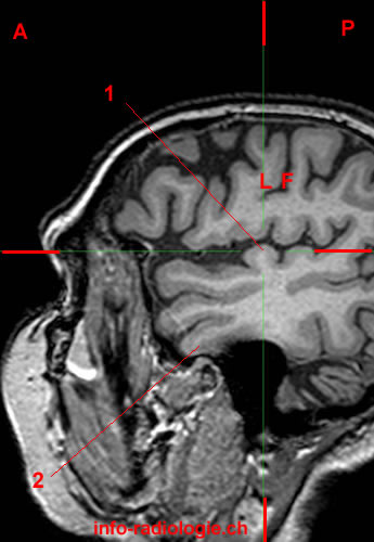
MRI of the brain, T1-weighted sagittal view. Level 2. Image 1.
1, Middle temporal gyrus. LF, Frontal lobe. A, Anterior P, Posterior
-
MRI of the brain, T1-weighted axial view. Level 2. Image 2.
1, Middle temporal gyrus. 2, Globe (right side). 3, Basilar artery.
-
MRI of the brain, T1-weighted coronal view. Level 2. Image 3.
1, Middle temporal gyrus. 2, Lateral ventricle. 3, Lateral sulcus.
-
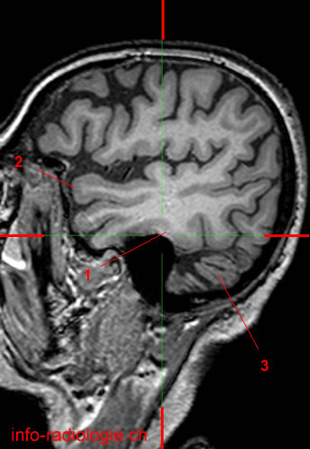
MRI of the brain, T1-weighted sagittal view. Level 3. Image 1.
1, Inferior temporal gyrus. LF, Frontal lobe. A, Anterior P, Posterior
-
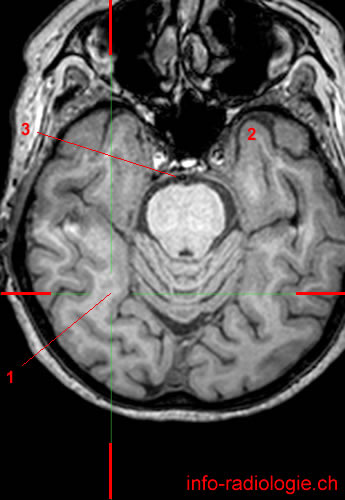
MRI of the brain, T1-weighted axial view. Level 3. Image 2.
1, Inferior temporal gyrus. 2, Maxillary sinus (right side). 3, Fourth ventricle.
-
MRI of the brain, T1-weighted coronal view. Level 3. Image 3.
1, Inferior temporal gyrus. 2, Superior frontal gyrus. 3, Lateral sulcus.
-
MRI of the brain, T1-weighted sagittal view. Level 4. Image 1.
1, gyri of Heschl. 2, Inferior temporal gyrus. LF, Frontal lobe. A, Anterior P, Posterior
-
MRI of the brain, T1-weighted axial view. Level 4. Image 2.
1, gyri of Heschl. 2, Globe (right side). 3, Lateral ventricle.
-
MRI of the brain, T1-weighted coronal view. Level 4. Image 3.
1, gyri of Heschl. 2, Lateral ventricle. 3, Cerebral peduncle.
-
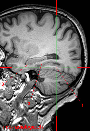
MRI of the brain, T1-weighted sagittal view. Level 5. Image 1.
1, Medial occipitotemporal gyrus. 2, Parahippocampal gyrus.3, Hippocampus.
-
MRI of the brain, T1-weighted axial view. Level 5. Image 2.
1, Medial occipitotemporal gyrus. 2, Temporal pole. 3, Basilar artery.
-
MRI of the brain, T1-weighted coronal view. Level 5. Image 3.
1, Medial occipitotemporal gyrus. 2, Calcarine sulcus. 3, Cingulate sulcus (pars marginal).
-
MRI of the brain, T1-weighted sagittal view. Level 6. Image 1.
1, Lateral occipitotemporal gyrus. 2, Superior temporal gyrus. 3, Cerebellum.
-
MRI of the brain, T1-weighted axial view. Level 6. Image 2.
1, Lateral occipitotemporal gyrus. 2, Maxillary sinus. 3, Pons. 4, Vermis.
-
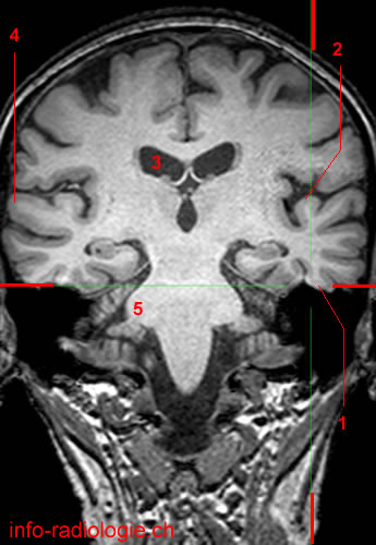
MRI of the brain, T1-weighted coronal view. Level 6. Image 3.
1, Lateral occipitotemporal gyrus. 2, gyri of Heschl. 3, Lateral ventricle. 4, Superior temporal gyrus. 5, Middle cerebellar peduncle.
Reference:
• Harnsberger HR, Osborn AG, Ross JS, Moore KR, Salzman KL, Carrasco CR, Halmiton BE, Davidson HC, Wiggins RH. Diagnostic and Surgical Imaging Anatomy: Brain, Head and Neck, Spine. 3rd ed. Salt Lake City, Utah. Amirsys. 2007.
• Bourjat P, Veillon F. Imagerie radiologique tête et cou. Paris, Vigot. 1995.
• Gouazé A, Baumann JA, Dhem A. Sobota. Atlas d'Anatomie humaine. Tome 3. Système nerveux central, système nerveux autonome, organe des sens et peau, vaisseaux et nerfs périphériques. 1er éd. Paris, Maloine. 1977.
• Kahle W, Cabrol C. Anatomie. Tome 3: Système nnerveux et organe des sens. 1er éd. Paris, Flammarion. 1979.
