Fornix
This photo gallery presents the anatomy of fornix by means of MRI (T1-weighted sagittal, axial and coronal views).
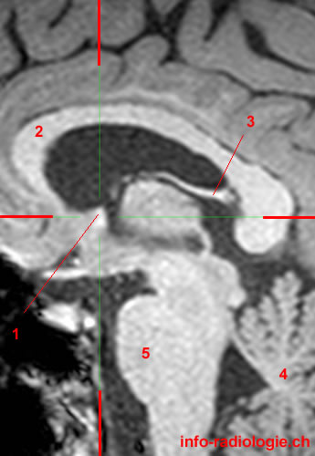
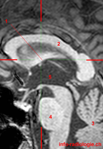
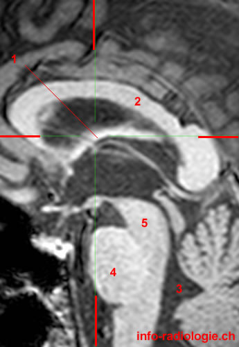
Brain MRI (magnification), T1-weighted, sagittal view. Level 1. Image 1.
1, Column of fornix. 2, Corpus callosum. 3, Crus of fornix. 4, Cerebellum. 5, Pons
-
Brain MRI (magnification), T1-weighted, sagittal view. Level 1. Image 1.
1, Column of fornix. 2, Corpus callosum. 3, Crus of fornix. 4, Cerebellum. 5, Pons
-
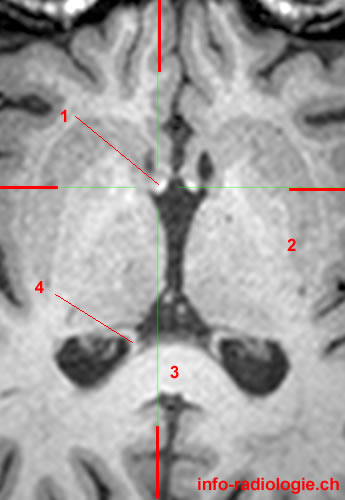
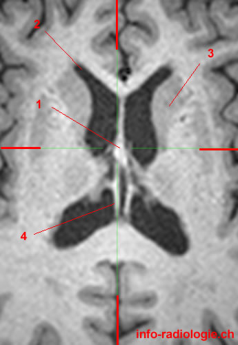
Brain MRI (magnification), T1-weighted, axial view. Level 1. Image 2.
1, Column of fornix. 2, Lentiform nucleus. 3, Corpus callosum. 4, Crus of fornix.
-
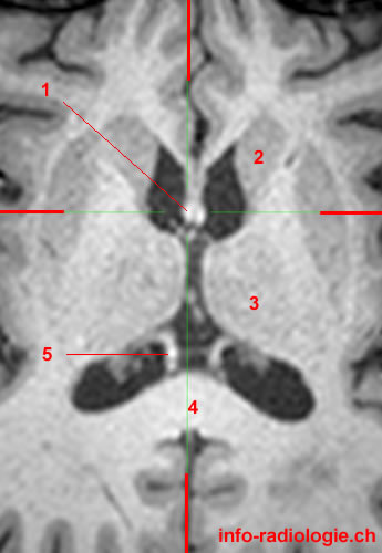
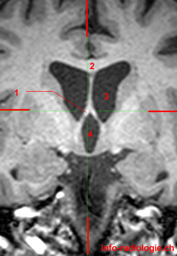
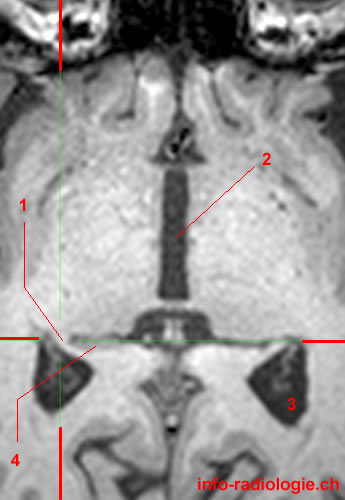
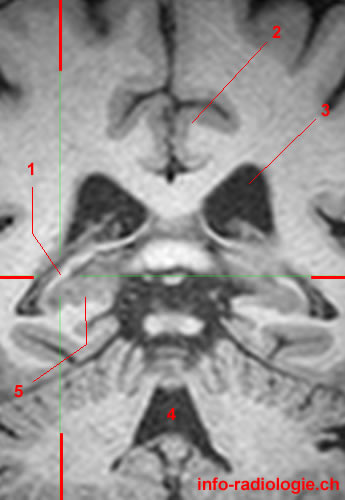
Brain MRI (magnification), T1-weighted, coronal view. Level 1. Image 3.
1, Column of fornix. 2, Corpus callosum. 3, Caudatus nucleus. 4, Hippocampus.
-
Brain MRI (magnification), T1-weighted, sagittal view. Level 2. Image 1.
1, Column of fornix. 2, Corpus callosum. 3, Midbrain. 4, Pons 5, Cerebellum.
-
Brain MRI (magnification), T1-weighted, axial view. Level 2. Image 2.
1, Column of fornix. 2, Caudatus nucleus. 3, Thalamus. 4, Corpus callosum. 5, Crus of fornix.
-
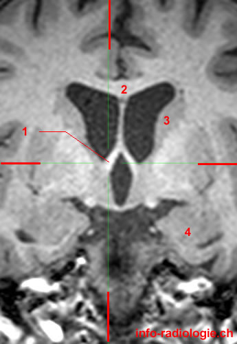
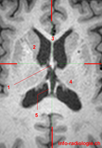
Brain MRI (magnification), T1-weighted, coronal view. Level 2. Image 3.
1, Column of fornix. 2, Corpus callosum. 3, Lateral ventricle. 4, Third ventricle.
-
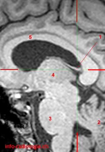
Brain MRI (magnification), T1-weighted, sagittal view. Level 3. Image 1.
1, Body of fornix. 2, Corpus callosum. 3, Cerebellum. 4, Pons 5, Third ventricle.
-
Brain MRI (magnification), T1-weighted, axial view. Level 3. Image 2.
1, Body of fornix. 2, Caudatus nucleus. 3, Corpus callosum. 4, Thalamus. 5, Crus of fornix.
-
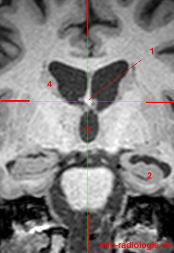
Brain MRI (magnification), T1-weighted, coronal view. Level 3. Image 3.
1, Body of fornix. 2, Hippocampus. 3, Third ventricle. 4, Caudatus nucleus.
-
Brain MRI (magnification), T1-weighted, sagittal view. Level 4. Image 1.
1, Body of fornix. 2, Corpus callosum. 3, Fourth ventricle. 4, Pons 5, Midbrain.
-
Brain MRI (magnification), T1-weighted, axial view. Level 4. Image 2.
1, Body of fornix. 2, Lateral ventricle. 3, Caudatus nucleus. 4, Crus of fornix.
-
Brain MRI (magnification), T1-weighted, coronal view. Level 4. Image 3.
1, Body of fornix. 2, Corpus callosum. 3, Lateral ventricle. 4, Third ventricle. 5, Hippocampus.
-
Brain MRI (magnification), T1-weighted, sagittal view. Level 5. Image 1.
1, Crus of fornix. 2, Cerebellum. 3, Pons 4, Thalamus. 5, Corpus callosum.
-
Brain MRI (magnification), T1-weighted, axial view. Level 5. Image 2.
1, Crus of fornix.
2, Column of fornix.
3, Lentiform nucleus.
-
Brain MRI (magnification), T1-weighted, coronal view. Level 5. Image 3.
1, Crus of fornix. 2, Cingulate gyrus. 3, Lateral ventricle. 4, Fourth ventricle.
-
Brain MRI (magnification), T1-weighted, sagittal view. Level 6. Image 1.
1, Crus of fornix.
2, Cerebellum.
3, Hippocampus.
4, Putamen.
-
Brain MRI (magnification), T1-weighted, axial view. Level 6. Image 2.
1, Crus of fornix. 2, Third ventricle. 3, Atrium. 4, Hippocampus.
-
Brain MRI (magnification), T1-weighted, coronal view. Level 6. Image 3.
1, Crus of fornix. 2, Cingulate gyrus. 3, Lateral ventricle. 4, Fourth ventricle.
Reference:
• Harnsberger HR, Osborn AG, Ross JS, Moore KR, Salzman KL, Carrasco CR, Halmiton BE, Davidson HC, Wiggins RH. Diagnostic and Surgical Imaging Anatomy: Brain, Head and Neck, Spine. 3rd ed. Salt Lake City, Utah. Amirsys. 2007.
• Bourjat P, Veillon F. Imagerie radiologique tête et cou. Paris, Vigot. 1995.
• Gouazé A, Baumann JA, Dhem A. Sobota. Atlas d'Anatomie humaine. Tome 3. Système nerveux central, système nerveux autonome, organe des sens et peau, vaisseaux et nerfs périphériques. 1er éd. Paris, Maloine. 1977.
• Kahle W, Cabrol C. Anatomie. Tome 3: Système nerveux et organe des sens. 1er éd. Paris, Flammarion. 1979.