Occipital Lobe
The occipital lobe is located in the posterior part of the cerebral hemispheres:
• posterior to the temporal lobe and parietal lobe
• the occipital lobe is partially separated from the parietal lobe by the parietooccipital fissure.
• In addition, there is only virtual separation between the temporal lobe and occipital lobe.
MRI of the brain, T1-weighted sagittal view. Level 1. Image 1.
1, Gyrus lingual. 2, Calcarine sulcus. 3, Parahippocampal gyrus. 4, Cerebellum.
-
MRI of the brain, T1-weighted sagittal view. Level 1. Image 1.
1, Gyrus lingual. 2, Calcarine sulcus. 3, Parahippocampal gyrus. 4, Cerebellum.
-
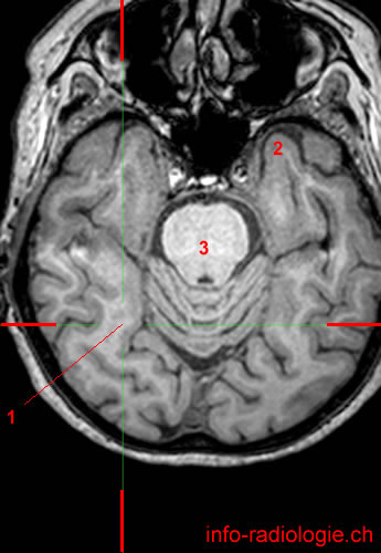
MRI of the brain, T1-weighted axial view. Level 1. Image 2.
1, Gyrus lingual. 2, Calcarine sulcus. 3, Cerebral aqueduct. 4, Temporal horn, lateral ventricle.
-
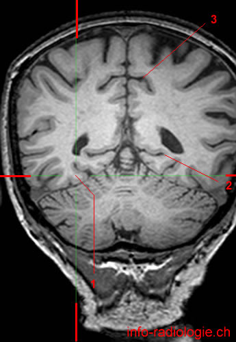
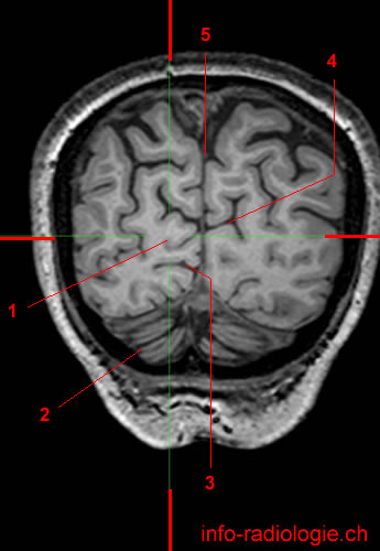
MRI of the brain, T1-weighted coronal view. Level 1. Image 3.
1, Gyrus lingual. 2, Calcarine sulcus. 3, Subparietal sulcus. 4, Cingulate sulcus (pars marginal).
-
MRI of the brain, T1-weighted sagittal view. Level 2. Image 1.
1, Medial occipitotemporal gyrus. 2, Calcarine sulcus. 3, Central sulcus.
-
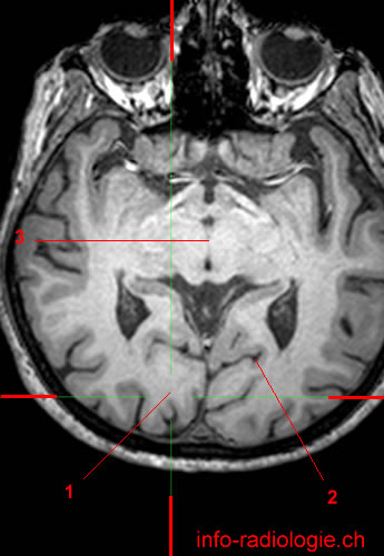
MRI of the brain, T1-weighted axial view. Level 2. Image 2.
1, Medial occipitotemporal gyrus. 2, Temporal lobe. 3, Pons.
-
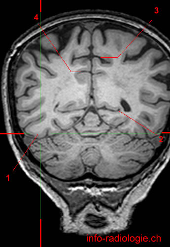
MRI of the brain, T1-weighted coronal view. Level 2. Image 3.
1, Medial occipitotemporal gyrus. 2, Calcarine sulcus. 3, Cingulate sulcus (pars marginal).
-
MRI of the brain, T1-weighted sagittal view. Level 3. Image 1.
1, Lateral occipitotemporal gyrus. 2, Cerebellum. 3, Lateral sulcus.
-
MRI of the brain, T1-weighted axial view. Level 3. Image 2.
1, Lateral occipitotemporal gyrus. 2, Superior cerebellar peduncle. 3, Temporal lobe (left side).
-
MRI of the brain, T1-weighted coronal view. Level 3. Image 3.
1, Lateral occipitotemporal gyrus. 2, Calcarine sulcus. 3, Cingulate sulcus (pars marginal). 4, Subparietal sulcus.
-
MRI of the brain, T1-weighted sagittal view. Level 4 (last level). Image 1.
1, Cuneus. 2, Parietooccipital sulcus. 3, Lateral ventricle. 4, Cerebellum. 5, Gyrus lingual. 6, Calcarine sulcus.
-
MRI of the brain, T1-weighted axial view. Level 4 (last level). Image 2.
1, Cuneus. 2, Parietooccipital sulcus. 3, Midbrain.
-
MRI of the brain, T1-weighted coronal view. Level 4 (last level). Image 3.
1, Cuneus. 2, Cerebellum. 3, Calcarine sulcus. 4, Parietooccipital sulcus. 5, Interhemispheric fissure.
Reference
• Harnsberger HR, Osborn AG, Ross JS, Moore KR, Salzman KL, Carrasco CR, Halmiton BE, Davidson HC, Wiggins RH. Diagnostic and Surgical Imaging Anatomy: Brain, Head and Neck, Spine. 3rd ed. Salt Lake City, Utah. Amirsys. 2007.
• Bourjat P, Veillon F. Imagerie radiologique tête et cou. Paris, Vigot. 1995.
• Gouazé A, Baumann JA, Dhem A. Sobota. Atlas d'Anatomie humaine. Tome 3. Système nerveux central, système nerveux autonome, organe des sens et peau, vaisseaux et nerfs périphériques. 1er éd. Paris, Maloine. 1977.
• Kahle W, Cabrol C. Anatomie. Tome 3: Système nerveux et organe des sens. 1er éd. Paris, Flammarion. 1979.
