-
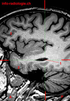
Level 1. Image 1. MRI of Brain (magnification), sagittal T1-weighted image. 1, Temporal horn of lateral ventricle. 2, Temporal lobe. 3, Frontal lobe. A, Anterior. P, Posterior.
-
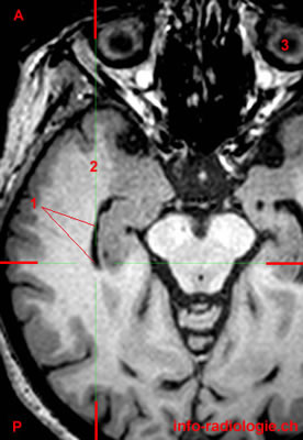
Level 1. Image 2. MRI of Brain (magnification), axial T1-weighted image. 1, Temporal horn of lateral ventricle. 2, Temporal lobe. 3, Globe. A, Anterior. P, Posterior.
-
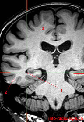
Level 1. Image 3. MRI of Brain (magnification), coronall T1-weighted image. 1, Temporal horn of lateral ventricle. 2, Temporal lobe. 3, Frontal lobe.
-
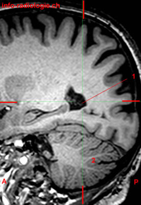
Level 2. Image 1. MRI of Brain (magnification), sagittal T1-weighted image. 1, Atrium (lateral ventricle). 2, Cerebellum. A, Anterior. P, Posterior.
-
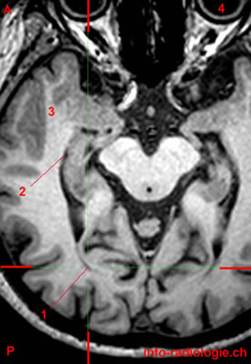
Level 2. Image 2. MRI of Brain (magnification), axial T1-weighted image. 1, Atrium (lateral ventricle). 2, Temporal lobe. A, Anterior. P, Posterior.
-
Level 2. Image 3. MRI of Brain (magnification), coronall T1-weighted image. 1, Atrium (lateral ventricle). 2, Cerebellum.
-
Level 3. Image 1. MRI of Brain (magnification), sagittal T1-weighted image. 1, Occipital horn of lateral ventricle. 2, Cerebellum. 3, Nucleus caudatus. A, Anterior. P, Posterior.
-
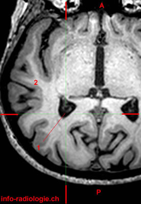
Level 3. Image 2. MRI of Brain (magnification), axial T1-weighted image. 1, Occipital horn of lateral ventricle. 2, Temporal horn of lateral ventricle. 3, Temporal lobe (right side). 4, Globe. A, Anterior. P, Posterior.
-
Level 3. Image 3. MRI of Brain (magnification), coronall T1-weighted image. 1, Occipital horn of lateral ventricle. 2, Cerebellum.
-
Level 4. Image 1. MRI of Brain (magnification), sagittal T1-weighted image. 1, Body of lateral ventricle. 2, Nucleus caudatus. 3, Cerebellum. A, Anterior. P, Posterior.
-
Level 4. Image 2. MRI of Brain (magnification), axial T1-weighted image. 1, Body of lateral ventricle. 2, Nucleus caudatus. A, Anterior. P, Posterior.
-
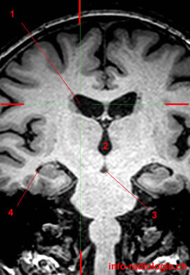
Level 4. Image 3. MRI of Brain (magnification), coronall T1-weighted image. 1, Body of lateral ventricle. 2, Third ventricle. 3, Interpeduncular cistern. 4, Temporal horn of lateral ventricle.
-
Level 5 (last level). Image 1. MRI of Brain (magnification), sagittal T1-weighted image. 1, Frontal horn of lateral ventricle. 2, Nucleus caudatus. 3, Cerebellum. A, Anterior. P, Posterior.
-
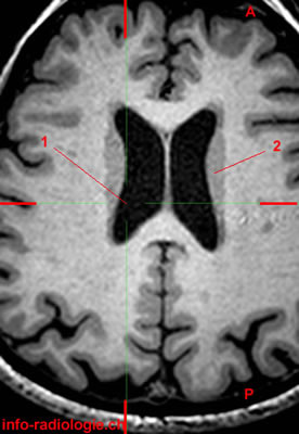
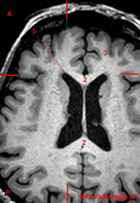
Level 5 (last level). Image 2. MRI of Brain (magnification), axial T1-weighted image. 1, Frontal horn of lateral ventricle. 2, Corpus callosum. 3, Frontal lobe. A, Anterior. P, Posterior.
-
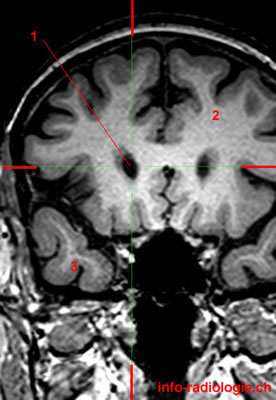
Level 5 (last level). Image 3. MRI of Brain (magnification), coronal T1-weighted image. 1, Frontal horn of lateral ventricle. 2, Frontal lobe gauche. 3, Temporal lobe (right side).