-
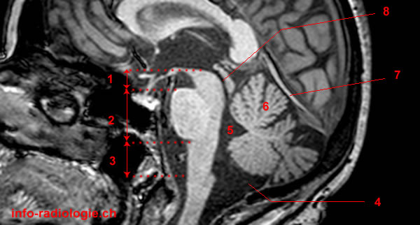
MRI of the brain, T1-weighted sagittal view. 1, Midbrain. 2, Pons. 3, Medulla. 4, Cisterna magna. 5, Fourth ventricle. 6, Cerebellum. 7, Tentorium cerebelli. 8, Cerebral aqueduct (of Sylvius).
-
MRI of the brain, T1-weighted coronal view. 1, Midbrain. 2, Pons. 3, Medulla. 4, Lateral ventricle. 5, Third ventricle. 6, Interpeduncular cisterna. 7, Temporal lobe. 8, Cervical cord.
-
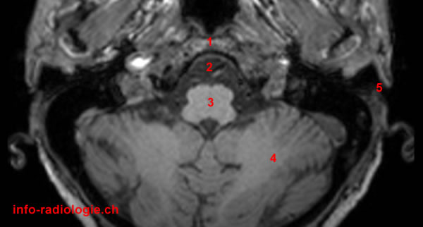
MRI of the brain, T1-weighted axial view. 1, Clivus. 2, Premedullary cistern. 3, Medulla. 4, Cerebellum. 5, External auditory canal.
-
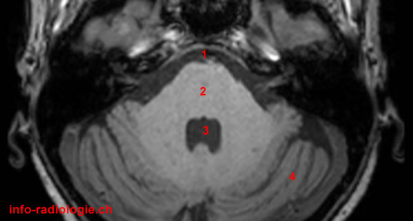
MRI of the brain, T1-weighted axial view. 1, Prepontine cisterna. 2, Pons. 3, Fourth ventricle. 4, Cerebellum.
-
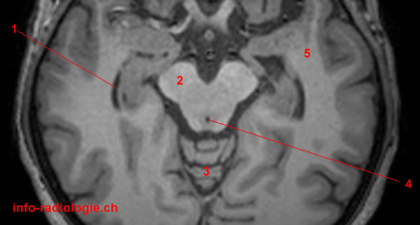
MRI of the brain, T1-weighted axial view. 1, Temporal lobe (right side). 2, Pons. 3, Vermis. 4, Fourth ventricle. 5, Temporal lobe (left side).
-
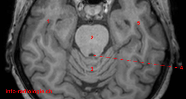
MRI of the brain, T1-weighted axial view. 1, Temporal horn of lateral ventricle (right side). 2, Cerebral peduncle. 3, Vermis. 4, Cerebral aqueduct (of Sylvius). 5, Temporal lobe (left side).