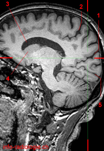
Parietooccipital sulcus separates partially the parietal lobe from occipital lobe in his medial part. Parietooccipital sulcus delimits the precuneus (parietal lobe) from the cuneus (occipital lobe).
Image 1. MRI of the brain, T1-weighted sagittal view.
1, Parietooccipital sulcus. 2, Cingulate sulcus (Pars marginal). 3, Lateral ventricle. 4, Caudate nucleus. 5, Calcarine sulcus.
• Harnsberger HR, Osborn AG, Ross JS, Moore KR, Salzman KL, Carrasco CR, Halmiton BE, Davidson HC, Wiggins RH. Diagnostic and Surgical Imaging Anatomy: Brain, Head and Neck, Spine. 3rd ed. Salt Lake City, Utah. Amirsys. 2007.
• Bourjat P, Veillon F. Imagerie radiologique tête et cou. Paris, Vigot. 1995.
• Gouazé A, Baumann JA, Dhem A. Sobota. Atlas d'Anatomie humaine. Tome 3. Système nerveux central, système nerveux autonome, organe des sens et peau, vaisseaux et nerfs périphériques. 1er éd. Paris, Maloine. 1977.
• Kahle W, Cabrol C. Anatomie. Tome 3: Système nnerveux et organe des sens. 1er éd. Paris, Flammarion. 1979.