-
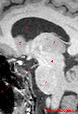
MRI of the brain, T1-weighted sagittal view. Image 1. 1, Lateral ventricle. 2, Caudatus nucleus. 3, Thalamus. 4, Midbrain. 5, Oculomotor nerve (III). 6, Pons.
-
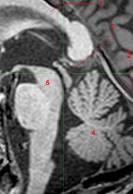
MRI of the brain, T1-weighted sagittal view. Image 2. 1, Lateral ventricle. 2, Caudatus nucleus. 3, Midbrain. 4, Cerebellar hemisphere.
-
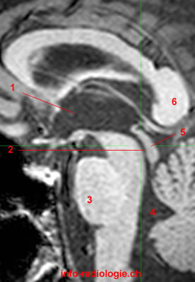
MRI of the brain, T1-weighted sagittal view. Image 3. 1, Third ventricle. 2, Cerebral aqueduct (of Sylvius). 3, Pons. 4, Fourth ventricle. 5, Tectum. 6, Splenium, Corpus callosum.
-
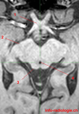
MRI of the brain, T1-weighted axial view. Image 4. 1, Optic chiasm. 2, Cerebral peduncle. 3, Superior colliculus. 4, Cerebral aqueduct (of Sylvius).
-
MRI of the brain, T1-weighted axial view. Image 5. 1, Substantia nigra. 2, Tegmentum. 3, Tectum. 4, Cerebral peduncle
-
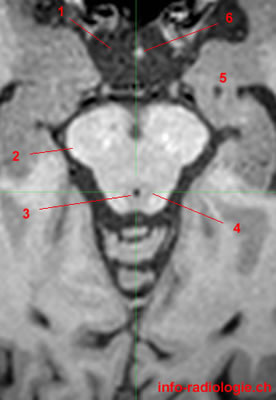
MRI of the brain, T1-weighted axial view. Image 6. 1, Suprasellar cistern. 2, Cerebral peduncle. 3, Cerebral aqueduct (of Sylvius). 4, Periaqueductal grey matter. 5, Hippocampus. 6, Pituitary stalk.
-
MRI of the brain, T1-weighted coronal view. Image 7. 1, Superior colliculus. 2, Fourth ventricle. 3, Inferior colliculus. 4, Lateral ventricle. 5, Corpus callosum.
-
MRI of the brain, T1-weighted coronal view. Image 8. 1, Hippocampus. 2, Cerebellar hemisphere. 3, Fourth ventricle. 4, Cerebral aqueduct (of Sylvius). 5, Lateral ventricle.
-
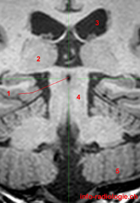
MRI of the brain, T1-weighted coronal view. Image 9 of 9. 1, Cerebral aqueduct (of Sylvius). 2, Thalamus. 3, Lateral ventricle. 4, Tegmentum. 5, Cerebellar hemisphere.