-
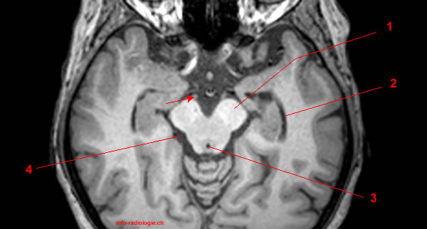
MRI of brain, axial T1-weighted image. Image 1.
1, Orbital cavity. 2, Temporal pole (right side). 3, Interpeduncular cistern. Arrow, Emergence of oculomotor nerve.
-
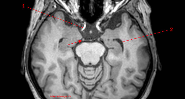
MRI of brain, axial T1-weighted image. Image 2.
1, Cerebral peduncle. 2, Temporal horn of lateral ventricle. 3, Cerebral aqueduct. 4, Ambient cistern. Arrow, Oculomotor nerve.
-
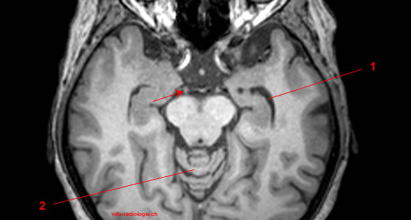
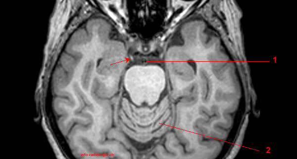
MRI of brain, axial T1-weighted image. Image 3.
1, Temporal horn of lateral ventricle. 2, Vermis. Arrow, Oculomotor nerve.
-
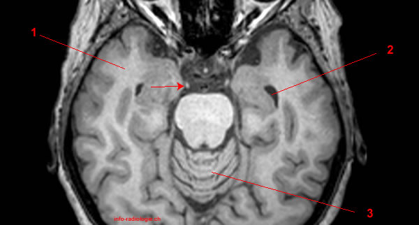
MRI of brain, axial T1-weighted image. Image 4.
1, Internal carotid artery (right side). 2, Temporal horn of lateral ventricle. Arrow, Oculomotor nerve.
-
MRI of brain, axial T1-weighted image. Image 5.
1, Temporal pole (right side). 2, Temporal pole (left side). Arrow, Oculomotor nerve.
-
MRI of brain, axial T1-weighted image. Image 6.
1, Temporal pole (right side). 2, Temporal horn of lateral ventricle. 3, Vermis. Arrow, Oculomotor nerve.
-
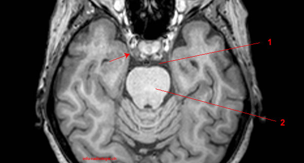
MRI of brain, axial T1-weighted image. Image 7.
1, Basilar artery. 2, Vermis. Arrow, Oculomotor nerve.
-
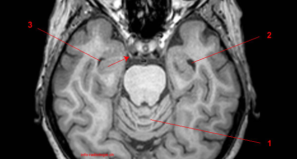
MRI of brain, axial T1-weighted image. Image 8.
1, Vermis. 2, Temporal horn of lateral ventricle gauche. 3, Temporal horn of lateral ventricle droit. Arrow, Oculomotor nerve.
-
MRI of brain, axial T1-weighted image. Image 9.
1, Prepontine cistern. 2, Pons. Arrow, Oculomotor nerve.
-
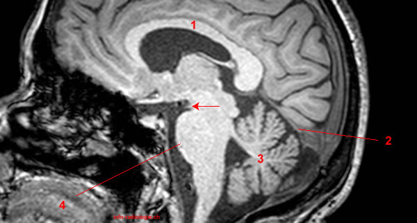
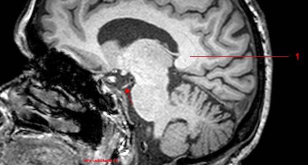
MRI of brain, sagittal T1-weighted image. Image 10.
1, Lateral ventricle. 2, Fourth ventricle. Arrow, Emergence of oculomotor nerve.
-
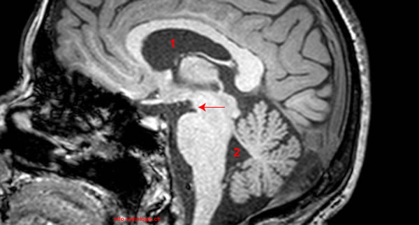
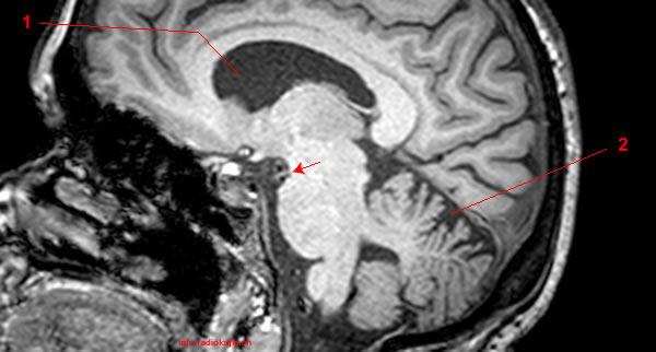
MRI of brain, sagittal T1-weighted image. Image 11.
1, Corpus callosum. 2, Cerebellar tentorium. 3, Cerebellum. 4, Pons. Arrow, Emergence of oculomotor nerve.
-
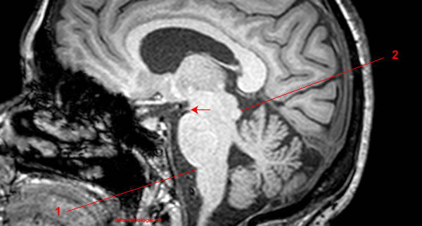
MRI of brain, sagittal T1-weighted image. Image 12.
1, Jonction Ponso-bulbaire. 2, Inferior colliculus. Arrow, Oculomotor nerve.
-
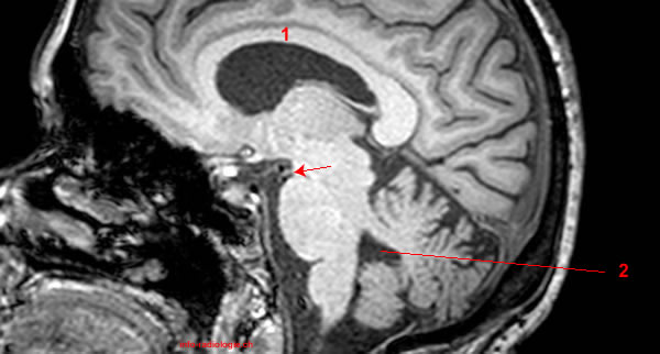
MRI of brain, sagittal T1-weighted image. Image 13.
1, Corpus callosum. 2, Fourth ventricle. Arrow, Oculomotor nerve.
-
MRI of brain, sagittal T1-weighted image. Image 14.
1, Lateral ventricle. 2, Superior cerebellar cistern. Arrow, Oculomotor nerve.
-
MRI of brain, sagittal T1-weighted image. Image 15.
1, Corpus callosum. 2, Lateral ventricle. 3, Quadrigeminal cistern. Arrow, Oculomotor nerve.
-
MRI of brain, sagittal T1-weighted image. Image 16.
1, Splenium of corpus callosum. Arrow, Oculomotor nerve.
-
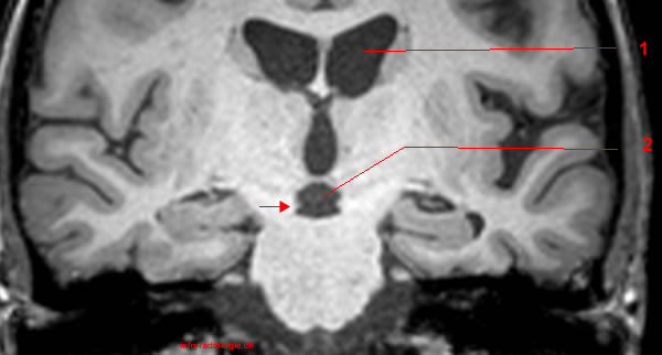
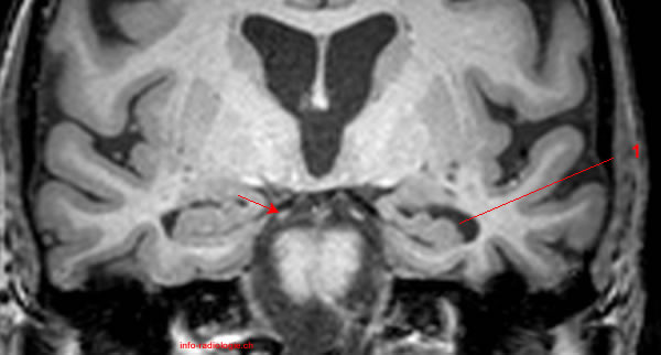
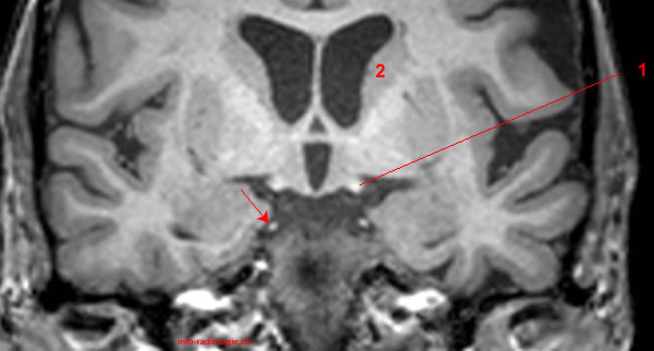
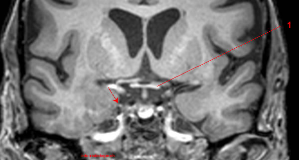
MRI of brain, coronal T1-weighted image. Image 17.
1, Lateral ventricle. 2, Interpeduncular cistern. Arrow, Emergence of oculomotor nerve.
-
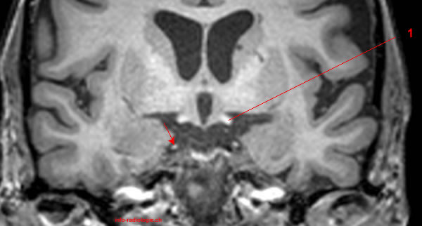
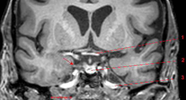
MRI of brain, coronal T1-weighted image. Image 18.
1, Septum pellucidum. 2, Third ventricle. Arrow, Emergence of oculomotor nerve.
-
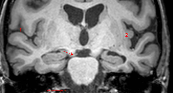
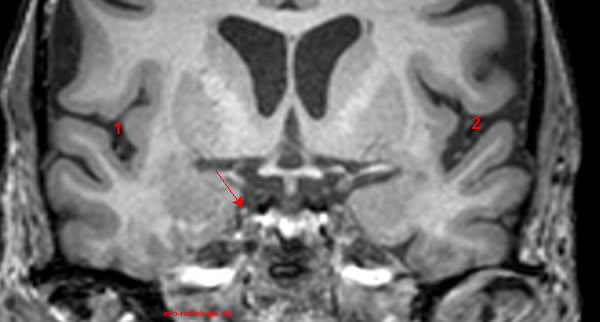
MRI of brain, coronal T1-weighted image. Image 19.
1, lateral sulcus (also called Sylvian fissure) . 2, Insula. Arrow, Oculomotor nerve.
-
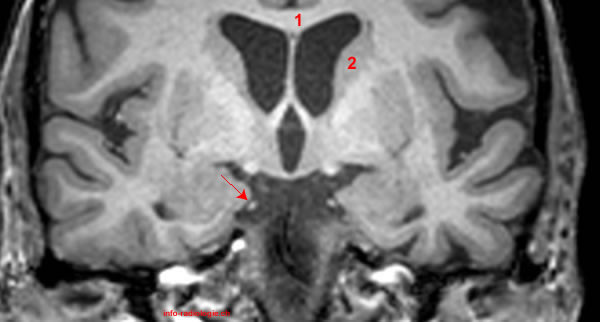
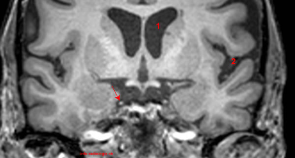
MRI of brain, coronal T1-weighted image. Image 20.
1, Lateral ventricle droit. 2, Lateral ventricle gauche. 3, Third ventricle. Arrow, Oculomotor nerve.
-
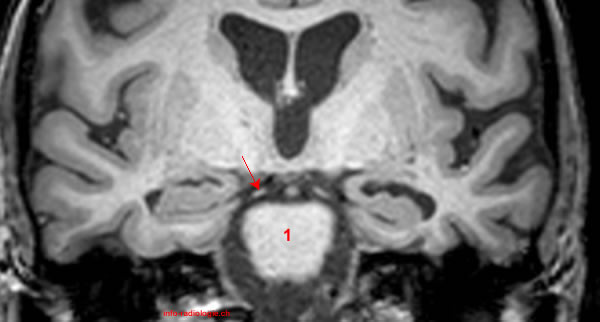
MRI of brain, coronal T1-weighted image. Image 21.
1, Pons. Arrow, Oculomotor nerve.
-
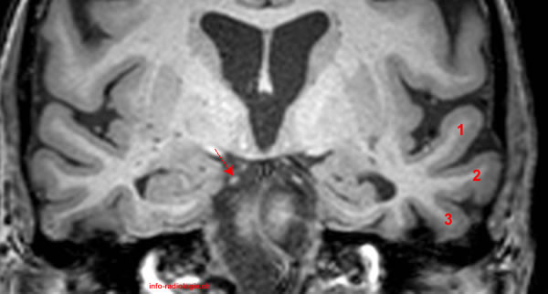
MRI of brain, coronal T1-weighted image. Image 22.
1, Temporal horn of lateral ventricle. Arrow, Oculomotor nerve.
-
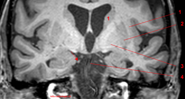
MRI of brain, coronal T1-weighted image. Image 23.
1, Superior temporal gyrus. 2, Middle temporal gyrus. 3, Inferior temporal gyrus. Arrow, Oculomotor nerve.
-
MRI of brain, coronal T1-weighted image. Image 24.
1, Claustrum. 2, Putamen. 3, Globus pallidus. 4, Basilar artery. Arrow, Oculomotor nerve.
-
MRI of brain, coronal T1-weighted image. Image 25.
1, Claustrum. 2, Putamen. 3, Globus pallidus. 4, Basilar artery. Arrow, Oculomotor nerve.
-
MRI of brain, coronal T1-weighted image. Image 26.
1, Corpus callosum. 2, Caudate nucleus. Arrow, Oculomotor nerve.
-
MRI of brain, coronal T1-weighted image. Image 27.
1, Optic tract. 2, Caudate nucleus. Arrow, Oculomotor nerve.
-
MRI of brain, coronal T1-weighted image. Image 28.
1, Optic tract. Arrow, Oculomotor nerve.
-
MRI of brain, coronal T1-weighted image. Image 29.
1, Lateral ventricle. 2, lateral sulcus (also called Sylvian fissure) (left side). Arrow, Oculomotor nerve.
-
MRI of brain, coronal T1-weighted image. Image 30.
1, lateral sulcus (also called Sylvian fissure) (right side). 2, lateral sulcus (also called Sylvian fissure) (left side). Arrow, Oculomotor nerve.
-
MRI of brain, coronal T1-weighted image. Image 31.
1, Optic chiasm. Arrow, Oculomotor nerve.
-
MRI of brain, coronal T1-weighted image. Image 32 de 32.
1, Optic chiasm. 2, Loge hypophysaire. 3, Internal carotid artery. Arrow, Oculomotor nerve.
