-
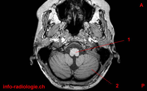
Brain MRI: axial cut, T1-weighted. Image 1.
1, Medulla. 2, Cerebellar hemisphere. A, Anterior. P, Posterior.
-
Brain MRI: axial cut, T1-weighted. Image 2.
1, Medulla. 2, Glossopharyngeal nerve (IX). 3, Cerebellar tonsil. 4, Cerebellar hemisphere. A, Anterior. P, Posterior.
-
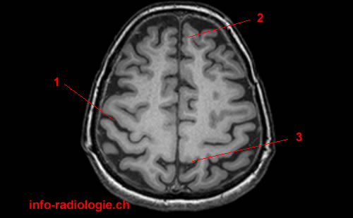
Brain MRI: axial cut, T1-weighted. Image 3.
1, Temporal lobe (Right side). 2, Pons. 3, Fourth ventricle 4, Cerebellar hemisphere. A, Anterior. P, Posterior.
-
Brain MRI: axial cut, T1-weighted. Image 4.
1, Vermis. 2, Basilar artery. 3, Globe (Right side). 4, Uncus. 5, Pons. A, Anterior. P, Posterior.
-
Brain MRI: axial cut, T1-weighted. Image 5.
1, Cerebral peduncle. 2, Middle cerebral artery. 3, Midbrain. 4, Cerebral aqueduct. A, Anterior. P, Posterior.
-
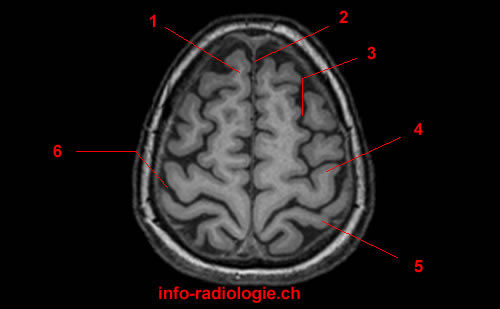
Brain MRI: axial cut, T1-weighted. Image 6.
1, Third ventricle. 2, Lateral sulcus. 3, Anterior commissure. 4, Pineal gland. 5, Choroid plexus (atrium of lateral ventricle). 6, Calcarine sulcus. A, Anterior. P, Posterior.
-
Brain MRI: axial cut, T1-weighted. Image 7.
1, Insula. 2, Septum pellucidum. 3, Genu of corpus callosum. 4, Caudate nucleus. 5, Anterior arm of internal capsule. 6, Putamen. 7, Thalamus. 8, pillars of the fornix A, Anterior. P, Posterior.
-
Brain MRI: axial cut, T1-weighted. Image 8.
1, Caudate nucleus. 2, Interhemispheric fissure. 3, Lateral ventricle. 4, Splenium of corpus callosum. A, Anterior. P, Posterior.
-
Brain MRI: axial cut, T1-weighted. Image 9.
1, Central sulcus 2, Superior frontal gyrus. 3, Interhemispheric fissure. A, Anterior. P, Posterior.
-
Brain MRI: axial cut, T1-weighted. Image 10.
1, Central sulcus 2, Interhemispheric fissure. 3, Cingulate sulcus (pars marginal). A, Anterior. P, Posterior.
-
Brain MRI: axial cut, T1-weighted. Image 11.
1, Superior frontal gyrus. 2, Interhemispheric fissure. 3, Superior frontal sulcus. 4, Precentral gyrus. 5, Postcentral gyrus. 6, Central sulcus. A, Anterior. P, Posterior.
-
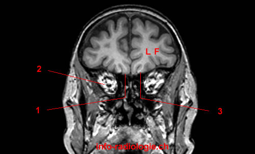
Image 12. Brain MRI: coronal cut, T1-weighted.1, Gyrus rectus. 2, Orbit. 3, Orbital gyrus. LF, Frontal lobe.
-
Image 13. Brain MRI: coronal cut, T1-weighted.1, Middle frontal gyrus. 2, Superior frontal gyrus. 3, Inferior frontal gyrus. 4, Temporal lobe.
-
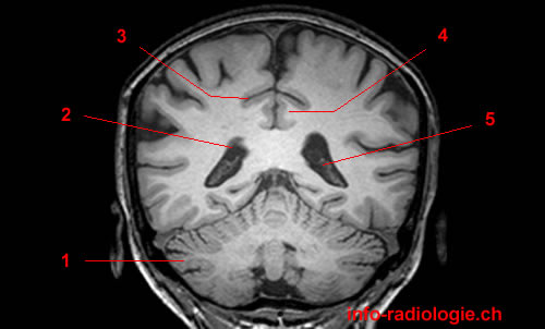
Image 14. Brain MRI: coronal cut, T1-weighted.1, Rostrum of corpus callosum. 2, Lateral ventricle. 3, Superior frontal gyrus. 4, Superior temporal gyrus. 5, Middle temporal gyrus.
-
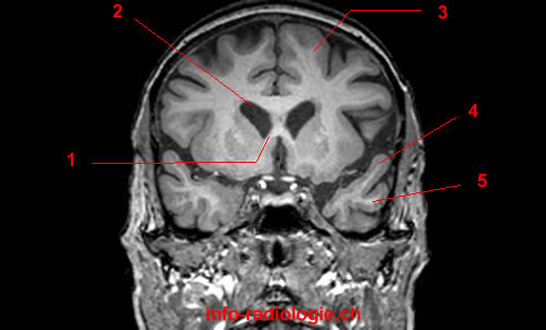
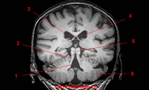
Image 15. Brain MRI: coronal cut, T1-weighted.1, Hippocampus. 2, Anterior commissure. 3, Column of fornix. 4, Lateral ventricle. 5, Lateral sulcus. 6, Middle temporal gyrus. 7, Inferior temporal gyrus.
-
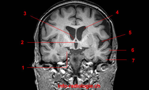
Image 16. Brain MRI: coronal cut, T1-weighted.1, Hippocampus. 2, Caudate nucleus. 3, Corpus callosum. 4, Lateral ventricle. 5, Third ventricle. 6, Interpeduncular cistern.
-
Image 17. Brain MRI: coronal cut, T1-weighted.1, Middle cerebellar peduncle. 2, Lateral sulcus. 3, Insula. 4, Fornix. 5, Cerebral peduncle.
-
Image 18. Brain MRI: coronal cut, T1-weighted.1, Fourth ventricle 2, Superior cerebellar peduncle. 3, Lateral ventricle. 4, Fornix. 5, Colliculus. 6, Cerebellar hemisphere.
-
Image 19. Brain MRI: coronal cut, T1-weighted.1, Cerebellar hemisphere. 2, Lateral ventricle. 3, Cingulate sulcus. 4, Cingulate gyrus. 5, Choroid plexus.
-
Image 20. Brain MRI: coronal cut, T1-weighted.1, Calcarine sulcus. 2, Subparietal sulcus. 3, Cingulate sulcus. 4, Cerebellar hemisphere.
-
Image 21. Brain MRI: coronal cut, T1-weighted.1, Parietooccipital sulcus. 2, Interhemispheric fissure. 3, Cerebellar hemisphere.
-
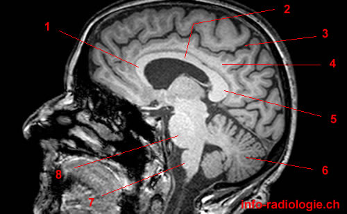
Image 22. Brain MRI: sagittal cut, T1-weighted.1, Genu of corpus callosum. 2, Trunk of corpus callosum. 3, Cingulate sulcus (pars marginal). 4, Cingulate gyrus. 5, Splenium of corpus callosum. 6, Cerebellum. 7, Medulla. 8, Pons.
-
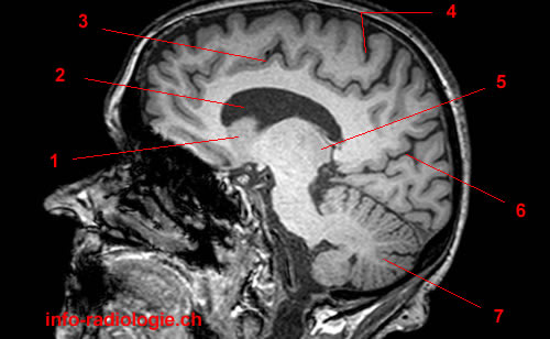
Image 23. Brain MRI: sagittal cut, T1-weighted.1, Caudate nucleus. 2, Lateral ventricle. 3, Cingulate sulcus. 4, Central sulcus 5, Thalamus. 6, Parietooccipital sulcus. 7, Cerebellum.
-
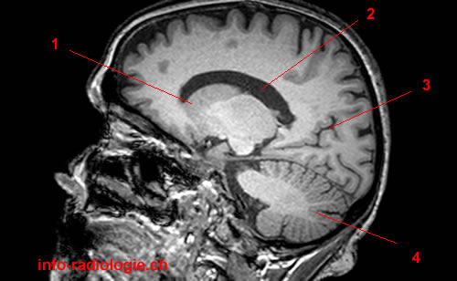
Image 24. Brain MRI: sagittal cut, T1-weighted.1, Caudate nucleus. 2, Lateral ventricle. 3, Parietooccipital sulcus. 4, Cerebellum.
-
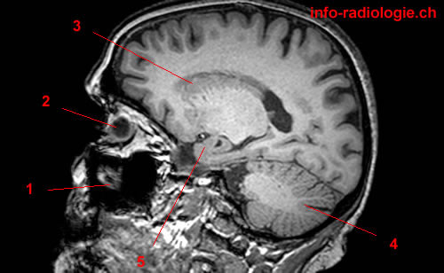
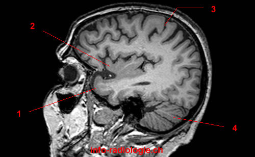
Image 25. Brain MRI: sagittal cut, T1-weighted.1, Maxillary sinus. 2, Globe. 3, Caudate nucleus. 4, Cerebellum. 5, Hippocampus.
-
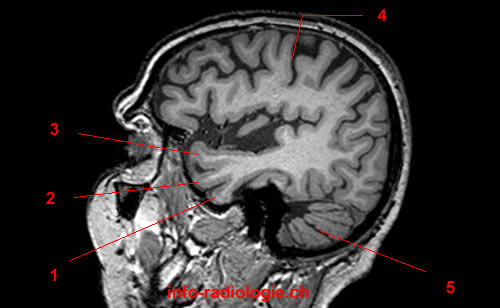
Image 26. Brain MRI: sagittal cut, T1-weighted.1, Gyrus parahippocampus. 2, Globe. 3, Caudate nucleus. 4, Atrium (lateral ventricle). 5, Gyrus lingual. 6, Medial occipitotemporal gyrus.
-
Image 27. Brain MRI: sagittal cut, T1-weighted.1, Hippocampus. 2, Central sulcus 3, Atrium (lateral ventricle). 4, Occipital pole. 5, Medial occipitotemporal gyrus.
-
Image 28. Brain MRI: sagittal cut, T1-weighted.1, Hippocampus. 2, Maxillary sinus. 3, Central sulcus 4, Cerebellum.
-
Image 29. Brain MRI: sagittal cut, T1-weighted.1, Temporal pole. 2, Insular cortex. 3, Central sulcus 4, Cerebellum.
-
Image 30. Brain MRI: sagittal cut, T1-weighted.1, Inferior temporal gyrus. 2, Middle temporal gyrus. 3, Superior temporal gyrus. 4, Central sulcus 5, Cerebellum.
-
Image 31 of 31. Brain MRI: sagittal cut, T1-weighted.1, Inferior temporal gyrus. 2, Middle temporal gyrus. 3, Superior temporal gyrus. 4, Lateral sulcus. 5, Central sulcus 6, Cerebellum.