Spinal cord
This webpage presents the anatomical structures found on spinal canal.
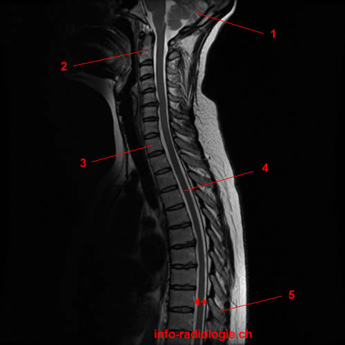
IRM cervico-dorsale, sagittal view, T2-weighted. Image 1.
1, Cerebellum . 2, C2 vertebra. 3, Vertèbre thoracique TH1. 4, Spinal cord. 5, Spinous process. Arrow, Spinal canal.
-
Cervicothoracic MRI, T2-weighted, sagittal view. Image 1.
1, Cerebellum . 2, C2 vertebra. 3, TH1 vertebra. 4, Spinal cord. 5, Spinous process. Arrow, Spinal canal.
-
Lumbar MRI, T2-weighted, sagittal view. Image 2.
1, L5 vertebra. 2, L1 vertebra. 3, Spinal cord. 4, Cerebrospinal fluid. 5, Nerve roots of cauda equina.
-
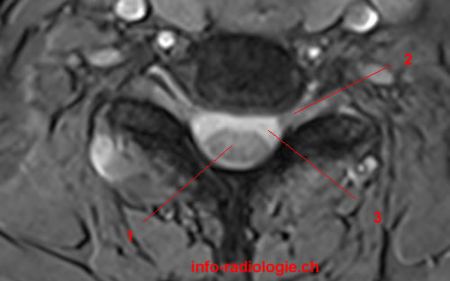
Cervical MRI scan, T2-weighted, axial view. Image 3.
1, Spinal cord. 2, Neural foramen. 3, Cerebrospinal fluid.
-
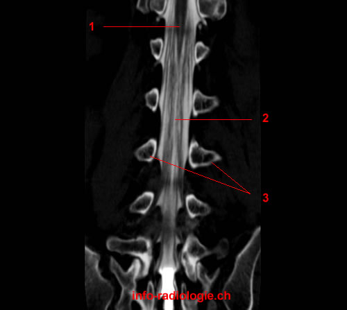
CT myelogram, lumbar spine, coronal view. Image 4.
1, Conus medullaris. 2, Nerve roots of cauda equina. 3, Pedicle.
-
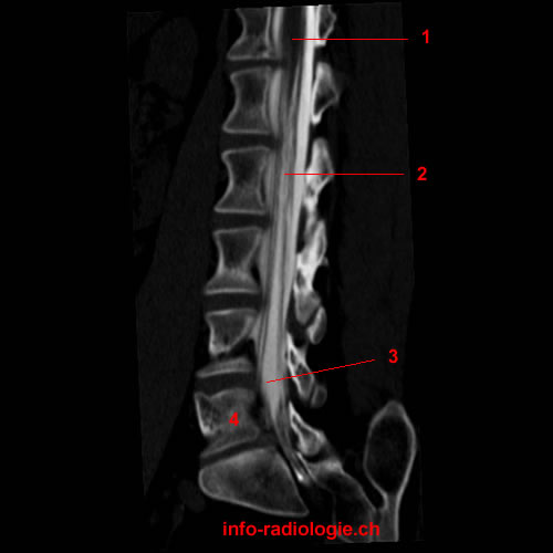
CT myelogram, lumbar spine, sagittal view. Image 5.
1, Conus medullaris. 2, Nerve roots of cauda equina. 3, Cerebrospinal fluid. 4, L5 Vertebral body.
-
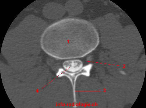
CT myelogram, lumbar spine, axial view. Image 6 of 6.
1, Vertebral body. 2, L4 nerve root / Neural foramen. 3, Spinous process. 4, Nerve roots of cauda equina.
Reference
• Harnsberger HR, Osborn AG, Ross JS, Moore KR, Salzman KL, Carrasco CR, Halmiton BE, Davidson HC, Wiggins RH. Diagnostic and Surgical Imaging Anatomy: Brain, Head and Neck, Spine. 3rd ed. Salt Lake City, Utah. Amirsys. 2007.
• Bourjat P, Veillon F. Imagerie radiologique tête et cou. Paris, Vigot. 1995.
• Gouazé A, Baumann JA, Dhem A. Sobota. Atlas d'Anatomie humaine. Tome 3. Système nerveux central, système nerveux autonome, organe des sens et peau, vaisseaux et nerfs périphériques. 1er éd. Paris, Maloine. 1977.
• Kahle W, Cabrol C. Anatomie. Tome 3: Système nnerveux et organe des sens. 1er éd. Paris, Flammarion. 1979.