The brain can be divided into left and right cerebral hemispheres.
Each cerebral hemisphere is divided into six sections, called "lobes": frontal lobe, parietal lobe, temporal and occipital lobe; plus two other that are not visible from outside: Insula and limbic lobe.
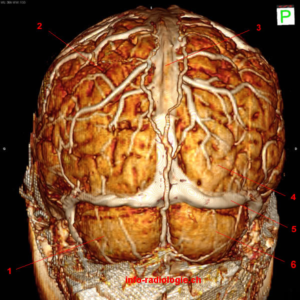
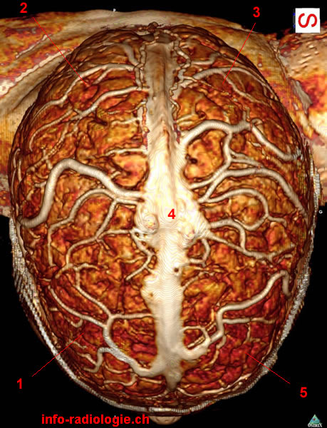
To locate these different lobes of the brain, a 3D reconstruction was made from a MRI exam of the head. The skull was removed using scissors provided by software (image processing).
Image 1. Brain, anterior view. 1, Frontal lobe (right side). 2, Superior sagittal sinus. 3, Frontal lobe (left side).

• Harnsberger HR, Osborn AG, Ross JS, Moore KR, Salzman KL, Carrasco CR, Halmiton BE, Davidson HC, Wiggins RH. Diagnostic and Surgical Imaging Anatomy: Brain, Head and Neck, Spine. 3rd ed. Salt Lake City, Utah. Amirsys. 2007.
• Bourjat P, Veillon F. Imagerie radiologique tête et cou. Paris, Vigot. 1995.
• Gouazé A, Baumann JA, Dhem A. Sobota. Atlas d'anatomie humaine. Tome 3. Système nerveux central, système nerveux autonome, organe des sens et peau, vaisseaux et nerfs périphériques. 1er éd. Paris, Maloine. 1977.
• Kahle W, Cabrol C. Anatomie. Tome 3: Système nerveux et organe des sens. 1er éd. Paris, Flammarion. 1979.